Category: Rad Tech Talk

Radiologic Technologist Blog

Effective Communication with Patients during Mammography

There are some basic guidelines for effective communication skills that X-ray techs and other healthcare workers can follow. These will allow you to establish a more effective relationship with your patients. The procedure of getting a mammography exam can be stressful for the patient, and their cooperation is important for achieving accurate results. Therefore, it is important to keep your patient calm and stress-free. This article will discuss how to effectively communicate with your patients to reduce their stress and get accurate imaging.

Continue reading

Mammography Certification for X-Ray Techs

You can work as a mammographer when you are a radiologic technologist, but you need to be certified. The mammography certification from ARRT is intended to ensure that you have the required knowledge to work with X-rays and handle the equipment carefully. To become a mammography technologist, you need at least an associate degree as a radiologic technologist, along with an ARRT certificate. In addition, most states require candidates to have specialized training to become a mammography technologist, and earn continuing education credits.

Continue reading

Radiation Exposure in Our Daily Life Compared to X-ray Exams

Several sources of radiation exposure are present in our environment. Radiation is an energy that travels through space at the speed of light. The energy comprises electric and magnetic fields, and possesses wave-like properties. Hence, radiation is also popular by the name of electromagnetic waves. They are present in two forms – ionizing and non-ionizing. Radiation is present in almost everything; some are harmful, while most are not.

There is a general conception that human beings experience radiation exposure only in X-ray rooms. However, this is not true. Things present in the environment also emit radioactivity that can be harmful. Radiation is harmful when the exposure is great and the dose exceeds human tolerance. For example, miners working in uranium mines are at a high risk of death because of excess radiation exposure. It could also result in long-term issues relating to health, such as cardiovascular disease or cancer. Read on to understand the exposure of radiation in our daily life as compared to X-ray exams.

Types of Radiation

There are two broad categories of radiation. They are further present in many forms based on their characteristics.

Non-Ionizing – Non-ionizing radiation is incapable of activating atoms or molecules because of the low energy levels. These are present in everyday products that surround us like microwaves, cellular telephones, cordless phones, remote control garage doors, etc., but in very low frequencies.

Ionizing – Ionizing radiation has high energy levels, which activate atoms by knocking out the electrons from their orbit. When the electron is moved, it creates an imbalance; thus, the atom receives a positive charge. These charged atoms and molecules are called ions, and the radiation that produces ions is called ionizing radiation.

Ionizing radiation is present in many forms:

Alpha Radiation

Alpha radiation has positively charged particles emitted by elements, such as uranium. It can enter the body through skin, easting, or breathing. A thin sheet of paper can be used to stop alpha radiation from entering the body. However, if it enters the body through food or air, it can cause biological damage to the body as the tissues would be directly exposed to the radiation.

Beta Radiation

Beta radiation has more penetrative properties than Alpha, and it can pass through water. The use of thick aluminum sheets or plastic sheets stops beta radiation from entering the body. However, it has the potential to induce damage to the exposed tissues.

Gamma Radiation

Gamma radiation has no charge or mass; thus, it has a deep penetrative property. It can pass through the human body if not stopped, and can cause major damage to the body as it passes. Dense materials like concrete absorb it.

Neutron Radiation

When neutrons interact with atoms of matter, they emit a type of radiation known as neutron radiation. The emission leads to a rise in other types of radiation. It is highly penetrative, and it can be absorbed by materials having a high amount of hydrogen atoms like plastics or paraffin wax, water, and concrete.

Natural Sources of Radiation Exposure

Radioactivity has existed from the beginning of time. Radioactive elements are present everywhere, from the earth’s crust to the walls of our house and the air we breathe. Humans are exposed to radioactive elements externally, but the human body also contains such elements in its muscles, bones, and tissues. Let us explore some other naturally occurring sources of radioactive elements.

Natural Gas

The gas used for cooking, heating, steaming, and other purposes exposes us to radioactive elements, but the exposure is minute; therefore, it is not harmful. The annual exposure to radiation through natural gas amounts to 9 mrem.

Radon

Soil and rocks emit a highly active, naturally occurring gas called radon. It is the major source of natural radiation exposure. It can get into houses via the floor or the walls. Radon is light enough to rise into the atmosphere. However, there is a great chance of it getting trapped inside the house and being inhaled. We can also ingest it through water or dust particles.

Air Travel

People working in the aviation industry who are frequent flyers like pilots, air hostesses, stewards, etc. are at a high risk of being endangered by harmful radiation. Aviation employees who fly regularly consume around 200 mrem annually as per the EPA.

Televisions, Microwaves, Cellphones

Human beings are exposed to radiation through radioactive elements that are emitted from televisions, microwave ovens, and other daily-use electrical items. But, the level of radiation exposure is too low to cause any damage. Did you know that more than 60 objects which we use in our daily lives expose us to harmful radiation?

X-ray Exposure

Exposure to X-rays occurs during medical procedures. Radiologic technologists conduct X-ray exams while wearing protective gear. They have control of the environment inside the examination room. The exposure to different examinations such as fluoroscopy, mammography, and CT scans differ from each other, and may even be high, but it is always kept under the dose limit set for patients.

Each body part reacts differently to radiation. Thus, the dose limit is different based on body parts. Radiation also has medical benefits; for example, radiation therapy is useful in curing cancer; however, a dose limit is set for all patients and workers in the radiologic department. The Environmental Protection Agency, Nuclear Regulatory Commission, and state agencies decide the dose limit.

Damage Caused by Exposure to X-rays in Comparison to Naturally Occurring Radiation

The exposure to naturally occurring radiation and X-rays does not affect the general public much. It is only harmful if radiation exposure is great. Workers in the radiology department and the nuclear testing department are at high risk of developing cancer even with limited tenure in these departments. People who work in uranium mines are at a high risk of inhaling radon, which could be fatal. Radon exposure in mines is much higher, and critically damages the exposed tissues.

Conclusion

With the increased use of radioactive elements in the coming future, it is important to look after people who work in places with various harmful radiation.

UIRP

Read more about this and other subjects and get 12 Category A ARRT® CE Credits in the X-Ray CE Course “ALARA and Radiation Protection”
Radiation Protection
Read More

Visit here to get information about radiation protection in x-ray imaging.

Requirement of Continuing Education Credits for MQSA Inspection

Continuing Education Credits are necessary for rad techs working in mammographic facilities. The CE credits help in qualifying the MQSA assessment. MQSA inspection is an annual affair. The inspectors certified under the MQSA evaluate the mammography facility. The assessment is based on the Mammography Quality Standard Act standards for facilities to provide proper patient care. In addition, the inspector informs the facility well in advance of the inspection date. The prior information helps the facility to adjust its schedule accordingly.


The evaluation covers everything present in the facility, such as:

  • The equipment
  • Services
  • Patient care
  • Certification
  • CE scores of the professionals working there

Every professional working there should have an active license that complies with the latest ARRT guidelines. This article will discuss how credits can help a facility pass the MQSA evaluation.

MQSA Inspection and CE Credits

MQSA evaluation inspectors follow guidelines set in 1999. To clarify, the main aim of conducting this annual inspection is to maintain mammography quality by providing the best quality services to patients around the U.S. The guidelines mentioned in the Act have an elongated description of the facility, and also includes credit requirements for people working there.

The following are some points you can use to prepare yourself for the inspection. Suppose the facility does not comply with the guidelines and quality standards set by the MQSA. In that case:

  • The license will nullify and the facility will no longer perform mammograms
  • The inspectors may also ban them from providing any service to patients

The facilities to undergo an evaluation need to have the following:

  • Every person should meet the quality standard set by the MQSA
  • The radiation dose should be under the permissible limit
  • The facility should have an accreditation certificate
  • The FDA authorizes agencies to certify such facilities
  • One should have a certification from the FDA to perform mammograms
  • The facility should display the certification visibly to mammography patients

Facilities must update the inspection certification. They should undergo MQSA inspection and evaluations by authorized physicists to maintain their certification. The facilities should take all the corrective actions when the inspectors point out a mistake.

Preparing for the evaluation

The annual evaluation is not only to assess the mammography units and their quality, but it also evaluates:

  • medical records
  • personnel and customer handling
  • medical audits

Below are the mentioned requirements of different departments during the MQSA assessment:

Requirements for personnel:

There are two requirements that workers have to comply with when working in a mammography service provider facility: the initial requirements, and the mammography continuing education requirements. Here, we will look at both of these requirements. The MQSA inspector will check these before marking you clear in the inspection.

Initial MQSA requirements:

You need to have one of the following with you:

  • State license for general radiography

OR

  • A general radiography certification from the FDA and the ARRT

All of the following points need to be checked:

  • 8 hours of training in all the mammographic modalities. These modalities should be the same that you will use while performing the mammographic exam.
  • You should have conducted 25 mammographic examinations under a MQSA-certified supervisor.
  • Subsequently, you should have 40 hours of training in mammography. The training should be documented and supervised by a qualified instructor. Your training must include training in
    • positioning and compression
    • quality control and assurance techniques
    • breast anatomy and physiology

Other than these, you must be trained in conducting mammography examinations on patients with breast implants.

MQSA requirements for individuals who qualified as radiologic technologists before 4/28/99 are a bit different. Look at the table below to find the information for yourself.

Qualified as a Radiologic Tech before 4/28/99 (INTERIM) Qualified as a Radiologic Tech after 4/28/99 (FINAL)

Must have one of the following:

Must have one of the following:

General Certification from the ARRT or the ARCRT ARRT General Certification
General Radiography License from any state Valid license from any state on General Radiography
Must have one of the following: Must have one of the following:
  • 40 hours of mammographic training
  • ARRT (M)
  • AZ Mammography Certification
  • NV Mammography Certification
  • CA Mammography Certification
  • Should have completed the FDA accepted training
40 hours of mammography training that must include the following:

  • Breast Anatomy
  • Positioning and Compression
  • Quality Control and Quality Assurance
  • Breast Implant mammography examination
  • Physiology
The MQSA allows attestation only if the training completion date is before 10/1/94 Should have conducted 25 exams under supervision
Start Date_____

(The Later of 10/1/94 or on the date when your last qualification was completed)

Start Date_____

(The Later of 10/1/94 or on the date when your last qualification was completed)

Should have 8 hours of training in any new modality if it has to be used. Should have 8 hours of training in any new modality if it has to be used.

These are the initial qualification requirements for personnel working in mammography facilities.  Most importantly, these need to be fulfilled as per their qualification completion date. In addition, all the information should be documented for the interviewer to check and verify.

Continuing mammography education credit requirements for mammography technologists

When you meet the initial requirements, you will conduct examinations under supervisors. You will need to earn continuing education credits; therefore, you will have permission to perform examinations of patients in the absence of a supervisor. You can earn these credits by taking a few courses or by teaching the modalities to others. You should earn the CE credits within 36 months immediately preceding the MQSA inspection date. Please bear the following points in mind:

  • Firstly, the credits you earn through teaching can be counted only once. For example, if you teach the course 4 times, the credits will only be counted once towards the 15 credit units.
  • Secondly, each modality should have six units; nothing less than that will be considered.

The new notice from the FDA Guidance Help System related to the 6 credit scores of mammography modalities suggests the following:

NOTE: The FDA is delaying enforcement of the six-hour new modality continuing education requirement indefinitely. Therefore, the inspector will assess the compliance with this requirement for each individual. It will be during the first facility inspection performed after the following date benchmarks have passed:
  • The third anniversary of the end date of the quarter calendar in which the candidate qualified for the initial requirements, or
  • June 30, 2002, or
  • The third-anniversary date when the candidate started using the new modality.

The FDA has intended to remove the requirement based on mammography modality continuing education credits on the recommendation of:

  • The National Mammography Quality Assurance Advisory Committee
  • The Institute of Medicine

Therefore, it has delayed the enforcement of the six-credit requirement for an indefinite period.

How will I know when my MQSA CE credits are due? 

You will need to check with your facility supervisor to know the MQSA inspection date. The inspector calculates the CE credits from the preceding 36 months from the inspection date. Some inspectors go with the quarterly reporting system. Therefore, only your facility supervisor will be able to tell you when your MQSA CE credits are due.

Where can I get additional information on MQSA CE requirements? 

The FDA requirements for MQSA inspection are duly displayed on the FDA’s Policy Guideline Help System for MQSA. Additionally, you will get answers to any query that you may have or need clarification on.

Can the application training that is received at work be counted towards my MQSA requirements? 

Yes, you can count the credits earned during application training at work towards the MQSA requirements. However, the credits may not apply to your state inspection requirements because each state inspection has different laws to comply with.

What documents should I present at the time of inspection? 

There are a few sets of documents that you need to present at the time of inspection. The documents are:

  • State license
  • Board certificate
  • Initial training requirement documentation
  • Certificate of 15 CE credits completion
  • A document containing information about you having completed the continuing experience requirement for the MQSA. Therefore, you should have experience with performing a minimum of 200 mammograms during the given time.

What if I fail to meet the MQSA CE requirements? 

Suppose you fail to meet the requirements of one or both of these:

  • continuing education credits
  • continuing experience credits

Consequently, you will have to hold performing non-supervised examinations. However, you will have 90 days to earn 15 continuing education credits from the date of inspection. After that, you can perform mammography examinations, but only under supervision. To complete your experience credits, you will have to perform 25 mammography examinations. However, these must be under the direct supervision of a MQSA-qualified radiologic technologist.

Final Words

The workers and equipment of the facility should meet the standards of the MQSA assessment. To clarify, workers need to earn continuing education credits per their position. Additionally, they have 36 months to earn the continuing education credits after starting their mammography practice individually. If a technologist fails to earn credits within the period, it may result in the dismissal of a practicing license. The MQSA evaluation ensures the well-functioning of the facility, resulting in improved patient care.

Visit here to get more information about arrt continuing education online.

Skull Fracture

The skull is made up of numerous flat bones that are connected by cranial sutures. Each flat bone has a thick outer table, a spongy diploe, and a narrower interior table. A thick, fibrous, adhesive dura mater lines the inner table. A narrow subdural gap exists between the dura’s inner surface and the thin arachnoid mater covering the brain.

Human Skull

Skull

A skull fracture is a break in the bone of the skull that usually develops as a consequence of direct trauma. Localized impact deforms the skull, which may cause harm to the cranial contents even if the skull does not fracture. Skull fractures are prevalent in both closed traumatic brain injury and penetrating traumatic brain injury. Their significance stems from the fact that they are related to a range of soft tissue injuries depending on their location.
A head injury and a skull fracture are not the same; rather, a skull fracture can be considered as part of a head injury. First, what exactly is a head injury?
A head injury is caused by damage to the scalp, skull, or brain. Head injuries are characterized as either closed (no cut or laceration to the skin) or penetrating (the skin and/or bone of the skull is fractured). Traumatic brain injuries range in severity from minor (referred to as mild traumatic brain injury) to severe (referred to as severe traumatic brain injury).
The symptoms of a head injury may appear shortly after the impact or they may appear gradually over several hours or days. The following are examples of specific types and symptoms of brain injuries:

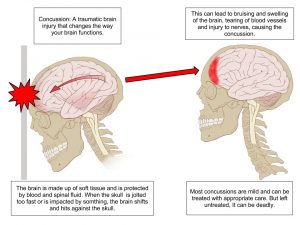
Concussion

Concussion of brain
Concussion

A concussion is sometimes known as a mild traumatic brain injury. This includes brain injuries produced by a blow to the head or body, a fall, or another type of trauma that rattles or shakes the brain inside the skull. Concussion victims may not always display symptoms that are visible to others. Concussions and mild head injuries can cause the following symptoms:
awareness loss ranging from a few seconds to a few minutes
consternation; memory and/or focus issues
dizziness
headache
Amnesia; memory loss of events occurring prior to or soon following the injury
vomiting and nausea
A changed state of awareness, such as drowsiness or difficulty waking up

Contusion

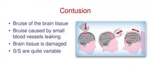
Contusion Bruise of the brain tissue

A bruise of the brain tissue is frequently linked with swelling (edema) and a rise in intracranial pressure (ICP). Dilated pupils are one of the symptoms of increased pressure within the brain and the skull. Other than that, the blood pressure could be too high, the patient may have a low pulse rate, irregular breathing, etc.

Fracture

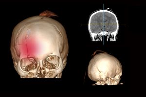
Brain with a fracture of the frontal part of the skull

A fracture is a break in the skull with or without a skin laceration. Tenderness is one of the symptoms of a skull fracture. Swelling, skull distortion, bruising around the eyes or behind the ear, and clear fluid pouring from the nose or ear are all symptoms of a skull fracture.

Bleeding

Brain Hemorrhage
Brain Hemorrhage

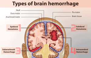
Bleeding in the brain, commonly known as a hemorrhage, is a potentially fatal disease that, in many situations, needs immediate care from a neurosurgeon. When a blood artery in the brain ruptures, it causes bleeding into surrounding tissue, edema, and increased intracranial pressure. Blood can also accumulate and form a clot, known as a hematoma. Clotting develops between the skull’s interior and the brain’s outer, robust membranous covering in an epidural hematoma (called the dura mater). A subdural hematoma is a collection of blood beneath the dura mater. The following symptoms of brain hemorrhage may start quickly or progressively worsen: sudden severe headache, convulsions nausea, or recurrent vomiting and lethargy, as well as loss of consciousness due to arm or leg weakness.

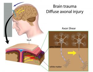
Shear Injury

Brain trauma with diffuse axon shear injury

Shear damage (also known as diffuse axonal injury) occurs when the brain collides violently with the inside of the skull. Nerve fibers that extend from the central body of a nerve cell are stretched or ripped, causing irreversible damage to brain cells and other issues within the nervous system. A shear injury’s principal symptom is a protracted loss of consciousness. Patients who display signs of brain injury should seek medical assistance immediately.
Head injuries are treated differently depending on the type of damage and the patient’s health. A physician may undertake a physical and neurologic exam, along with imaging studies to determine the severity of a head injury.

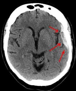
CT scan of the head :  CT scan of the head is performed. CT scanning combines specialized X-ray equipment with advanced computers to generate numerous images or photos of the head and brain. Doctors use CT scans of the head to identify bleeding, edema, brain damage, and skull fractures. More information regarding CT may be found on the Safety page.

Ct-scan of the brain

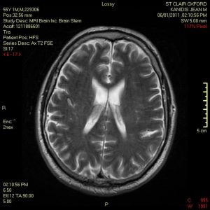
MRI of the brain: Magnetic Resonance Imaging (MRI) creates comprehensive images of organs, soft tissues, bone, and almost all other internal body components by using a high magnetic field, radiofrequency pulses, and a computer. In certain cases, clinicians will employ an MRI of the brain, including advanced MR scanning techniques such as diffusion-weighted imaging, diffusion tensor imaging, and MR spectroscopy, to help diagnose brain injuries that do not show up on a standard MR scan.

MRI of the brain
MRI of the brain

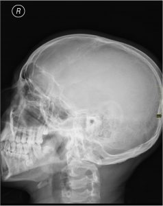
X-ray of the head: An X-ray is a procedure that includes exposing a portion of the body to a low dosage of ionizing radiation in order to obtain images of the inside of the body. Doctors occasionally use an X-ray of the head to discover and assess skull fractures. However, such an exam is rarely used nowadays because it cannot check the brain at all. A CT scan can show most skull fractures more clearly than a head X-ray.

X-ray of the head
X-ray of the head

Furthermore, clinicians clearly understand that it is more necessary to establish underlying brain damage in order to decide the right therapy for the patient. CT and, if necessary, MRI are the primary imaging methods for this purpose.
Skull fractures, on the other hand, can be generally classified as follows:
anatomically
base of skull
skull vault (calvaria)
associated with an overlying wound
open (compound)
closed
degree of displacement
undisplaced
depressed (5 to 10 mm)
number of fracture lines/fragments
linear
comminuted
Fractures of the skull, like fractures of any other bone, occur when biomechanical forces surpass the tolerance of the bone. The fracture pattern is determined by the position, direction, and kinematic aspects of the hit, along with intrinsic skull traits.
Radiographic features
Plain radiographs
Plain radiographs have a limited purpose, and are being phased out in favor of CT scans. They are no longer suggested for assessing head injuries unless they are part of a skeletal assessment for a suspected non-accidental child injury.
CT scan
A CT scan of the brain is the best way to visualize skull fractures. CT is not only sensitive to the diagnosis of fractures, but it may also describe their extent and assist in surgical planning. It is also collected at the same time that the brain is scanned.
A volumetric CT of the skull with tiny (1 mm) voxels that can be reconstructed in several planes should be obtained. If nondisplaced fractures are to be viewed, a bone algorithm must be utilized.
Fractures occur as discontinuities in the bone, and they may or may not be displaced. They must be recognized from normal sutures, which lack the corticated borders found in fractures. If the fracture affects a paranasal sinus, middle ear, or mastoid air cell, it will almost always contain some blood, which is a useful signal of the presence of an underlying fracture.
A thorough search for nearby soft tissue injuries should be performed when a fracture is found. Soft tissue injuries include the following:
Vascular:
arterial dissection, occlusion, or rupture
arterial extradural hemorrhage
arteriovenous fistula (e.g., direct carotid-cavernous fistula)
Dural venous sinus injury
venous extradural hemorrhage
Dural venous sinus thrombosis
extension through cranial nerve foramina or canals with neural damage
underlying cerebral hemorrhagic contusions
Dural tears leading to CSF leak and intracranial hypotension
MRI
MRI is insensitive to fractures, and it is frequently startling how difficult it is to image fractures even when they are obvious and well-documented on a CT.
Skull fractures that are closed and nondisplaced seldom require direct management, with therapy focusing on any related damage (e.g., extradural hematoma). Depressed fractures, on the other hand, frequently necessitate surgical intervention for cosmesis and a decrease in the frequency of post-traumatic epilepsy. Debridement is frequently required after an open (complex) fracture to limit the risk of infection.

Sources:
https://radiopaedia.org/articles/skull-fractures
https://www.radiologyinfo.org/en/info/headinjury

Get more information about radiologic technology continuing education and arrt ce credits online here.

Visit here to get more information about x-ray positioning.

Mammography Quality Standard Act Inspection Checklist

Before the Mammography Quality Standard Act was set up, there were many problems in the quality control process of mammographic equipment. Additionally, it was difficult to train mammography technologists to perform quality mammograms on patients, and inconsistencies in the system led to technicians performing suboptimal mammograms.

Therefore, to improve early breast cancer diagnosis, it was important to work on image resolution. Hence, the MQSA standards were set, and proper documentation was formed for everyone working in the mammography facility, including:

  • requirements for equipment
  • quality control
  • CE credits in both education and experience for mammography technologists, etc.

Certainly, these requirements are inspected by the MQSA inspector, and as a result, the facility is certified for providing quality mammography services to citizens. In this article, we will discuss the MQSA inspection checklist in detail to help you prepare for the evaluation day.

MQSA 1992

The Mammography Quality Standards Act (MQSA) was passed in 1992 by congress in order to achieve standard mammography quality across the nation. The decision was made to:

  • implement quality mammography services for patients
  • help in early breast cancer detection

The MQSA has been amended twice, taking into consideration the impact of setting up the requirement standards. Currently, the final amendment has detailed requirements for:

  • Employees: For example, the people who interpret mammograms, mammography techs, radio techs, physicists, etc. concerning their education, experience, and CE credits
  • Medical Audits: It is important for mammography facilities to have a system in order to follow up the obtained biopsy results
  • Quality Control and Recordkeeping: Facilities should always keep a record of their activities; certain records are required at the time of inspection

Above all, it is mandatory for facilities to get their MQSA certification in order to offer any mammography service to citizens. The facilities get certification only if they are able to comply with the MQSA requirements. If the facility fails to qualify the inspection, it has to stop providing all mammography services. Moreover, they have to regain their certificate by implementing the changes suggested by the inspector. Additionally, it is compulsory for facilities to display the valid certificate copy from the MQSA, and it must be easily visible to customers.

Who gets MQSA certified?

There is a general confusion that stays with radiologic and mammography technologists. You, as a mammography technologist or a radiologic technologist, do not get certified under the Mammography Quality Standard Act. The facility that you work in gets the certification to offer mammography services. Moreover, it is illegal to perform mammograms on people without a valid Mammography Quality Standard Act accreditation; however, it is mandatory for you as a tech to comply with the requirements set for you. You are responsible for the image resolution and comprehension of the mammograms; therefore, you will need to comply with the CE credits and the experience credits. Failure to fulfil these requirements will impact the certification for the facility.

The Inspection Checklist

The MQSA inspector informs the facility well in advance about the inspection date and time. The facility needs to prepare in such a way as to still be able to provide services to patients. The inspector will need approximately 20 minutes in each mammography unit to evaluate the mammogram. The following is a checklist for you to crosscheck your preparations for the inspection day.

Pre-Inspection requirements

  • Keep records of every activity that you perform
  • Document all the information
  • Keep ample space ready for the inspector to view the documents undisrupted

Reports

  • All other inspection reports that are conducted by the state within the last and the current MQSA inspection period
  • Safety Survey Reports of the equipment
  • Record retention policy
  • State registration documents upholding the present name, contact, and owner details
  • Availability of all the required protective shields for employees and equipment

Medical Records

  • Display all records; the inspector will select any 5 of his choice
  • Keep a medical record log that will help track positive results
  • Have letter samples to depict the use of easy language in result demonstration
  • Documentation to ensure that the physicians review all the reposts

Quality Control

  • EQUIP reports
  • Ensure that all the monitors and QC manuals are updated to the latest versions
  • Put forward all the QC manuals and QC tests of all the monitors (on-site and off-site) used since the last inspections
  • Ensure that the information is the same in the QC forms as it is in the one filed in the monitor software
  • Provide Medical Equipment Evaluation reports about all the units, new and old
  • Provide reports about the evaluation of the mamo unit after the replacement of any part.

Plan of Action Manual

  • Display the technique chart
  • Provide detailed information on the responsibilities associated with the role of every employee
  • Keep records of service and operator manuals of all mamo units
  • Keep physicist reports
  • Keep meeting minutes of the quality assurance meetings held at the facility to improve the service quality

Credential Requirements for Employees

On-site and Off-site Medical Physicists

Initial Qualifications

  • Degree certificate, 20 hours per semester in physics, 20 contact hours in mammography surveys
  • Should have survey 1 facility and 10 units or an FDA approval letter
  • Unexpired State Approval / License
  • 2-D and 3-D initial training – 8 CEU’s (only applicable if your facility uses the same)

Continuing Education / Experience Requirements

  • 15 CEUs in 3 years
  • Should have maintained a record of surveying at least 2 facilities and 6 units in 24 months

Radiologic Technologist

Initial Qualifications

  • Copy of the 40 hours of training and the ARRT R card
  • Have supervised 25 mammograms
  • 8 CE units of the 2-D and 3-D training (if used in your facility)

Continuing Education / Experience

Interpreting Physicians

Initial Qualifications

  • Copy of a valid MD license and ABR
  • 3 months of training and 60 CMEs (if qualified after 4/28/99)
  • 40 CMEs and 2 months of training
  • Should have supervised 240 readings within a period of 6 months
  • 8 CEUs in 2-D and 3-D initial training (only applicable if your facility uses them)

Continuing Education / Experience

  • Should have given readings of at least 960 mammograms in 2 years
  • Should have earned 15 Continuing Education Units in 3 years
  • Additionally, the CEUs must be in category 1

Conclusion

MQSA inspection is an annual affair, which aims to maintain a standard quality of mammography services around the country. The evaluation is complete only with the exit interview that the inspector gives. The inspector might inform you about the deficiencies during the exit interview or you may receive an email containing the result of the inspection.