Description
Get 12 X-Ray CE Credits now. Guaranteed**. Category A
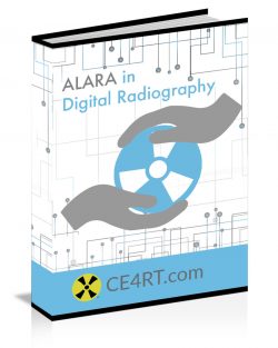
Is your patient dose As Low As Reasonably Achievable?
This mobile friendly continuing education course for Radiologic Technologists is a review of key concepts behind the science of ionizing radiation, and best practices in radiation protection for the working radiologic technologist.
Ionizing radiation is a potentially awesome force of nature. It surrounds us and effects us daily. It can be used to save lives. It also can become a weapon of mass destruction. As RTs we wield this awesome force and we need to understand it’s properties to do so safely. A better understanding can help us explain radiation to our patients, and to mitigate side effects imposed on those we intend to heal.
Although the fundamental principles of physics are discussed, this is not a physics course and will not require a calculator.
Delivered in layman’s terms with colorful illustrations, this course will walk you through definitions and explanations of our current understandings regarding ionizing radiation. It will explain atomic structure, charge, and ionization. It will also explain electromagnetic radiation, how x rays are formed, and how they interact with our patients bodies.
What really happens inside our patients on a molecular level when we take an X Ray? On a day to day basis, we don’t always stop and appreciate the physics behind what we do. Understanding the fundamental principles explained in this course will help to build a more solid understanding of what happens when we push the switch. Things that happen on an atomic level may seem insignificant. But multiplied by millions or billions of times they are a force to be respected and revered. Knowing this information will help you to explain to doctors and patients what is happening as well as give you tools to help alert you when things don’t go as expected. As a result, hopefully we can all be safer technologists.
CE Course Outline
Explaining Atoms, Particles, and Energy 
Electrons, Electrons Performing Work, Elementary Particles and Antiparticles, Ions, Atomic Number and Atomic Mass, Radionuclides, Electromagnetic Radiation, Photons, Photon Speed, Neutrons, Molecules
Types of Radiation
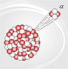 Electromagnetic Spectrum, Wavelength, Frequency, X-rays, UV, Gamma Rays, Radioactive Decay, Alpha Particles, Beta Particles, High vs. Low LET
Radiation Interacting With Matter
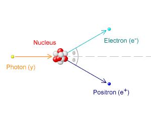
Shielding Radiation, Photoelectric Effect, Compton Effect, Pair Production, Annihilation Reaction, The Laws of Thermodynamics, Attenuation and Absorption, Half Value Layer
Measuring Energy
Basic Units of Measurement, Force, Acceleration, and Work, Mass, Einstein Mass and Energy, Friction and Resistance, Joules and Watts, Measuring Electrical Charge, Measuring Current
Sources of Radiation 
Sources of Radiation Exposure, Medical Procedures, Terrestrial Radiation, Cosmic Radiation, Actinides, Radon, Radon in Homes, Naturally Occurring Food Radiation, Nuclear fallout, Human Bodies, Depleted Uranium, Other Man Made Sources
Human Cell Biology
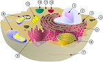 Nucleus and Organelles, Mitochondria, Ribosomes, Lysosomes, Endoplasmic reticulum, Golgi complex, Vacuoles, Cytoskeleton, DNA Chromosomes and Genes, Proteins, Enzymes, Hormones, Antibodies, Transcription, Translation, Mitosis
Biologic Effects of Ionizing Radiation
Dose Response, Tissue Radiosensitivity, Deterministic and Acute Effects, Stochastic Effects, DNA Damage, Gene Mutation, Proliferation of Genetic Damage, Radiation and Pregnancy, Nursing Mothers, Radiation and Cancer, Cataracts, Radiophobia
Medical Applications Using Radiation 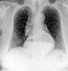
Medical X-rays, Computed Tomography, Nuclear Medicine, PET, Radiation Therapy, Radiosurgery
Monitoring Radiation and Exposure
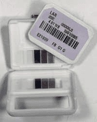 Dosimetry, Automated Dose Reporting, ACR’s Dose Index Registry, Fluoroscopy monitoring, Limitations of dose monitoring, Absorbed Dose, Equivalent Dose, Effective Dose, Committed Dose, Geiger Counters, Film Badges, Personal Dosimeters, Ionization Chambers, Whole Body Counter, Cytogenetic Biodosimetry
Radiation Protection for Staff
ALARA, Safety Controls, Time, Distance, and Shielding, Radiation Protection Agencies and Regulations, Radiation Protection for Radiographers, Scatter Radiation, Regulatory Limits for Occupational Exposure, Protective Apparel, Protection of Staff in Fluoroscopy, Pregnant Radiographers and Radiation Workers
Radiation Protection for Patients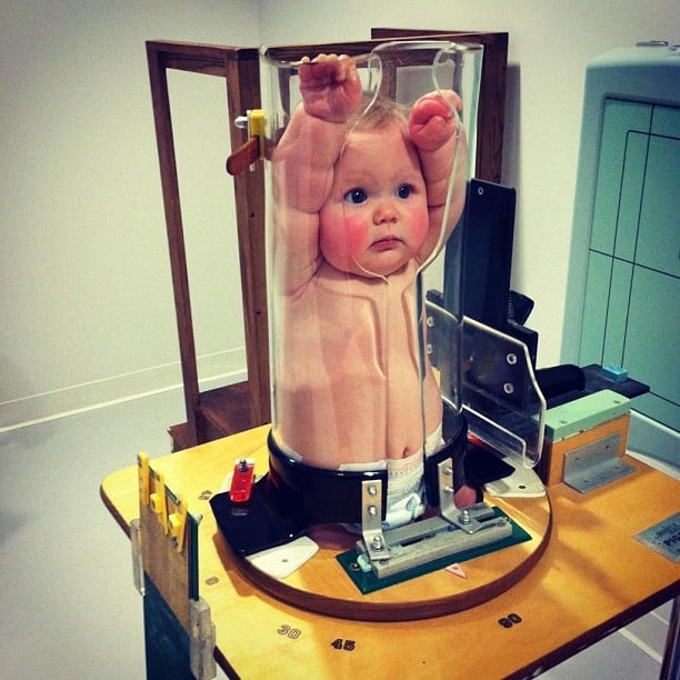
Avoiding Repeat Exposures, Communication, Exposure Technique Charts, Department Protocols, Filtration, Grids, Filters, and Air Gap, Screening for Pregnancy, High kVp Low mAs, Inverse Square Law, Digital and Computed Radiography, Exposure Indicators and Dose Creep, Collimation, Shielding, Pediatric Patients, Immobilization, Obese Patients, Stepping Lightly
ARRT®* STRUCTURED EDUCATION CREDIT DISTRIBUTION FOR THIS COURSE.
| DISCIPLINE | CATEGORY & SUBCATEGORIES | CE CREDITS PROVIDED |
|---|---|---|
| RAD – 2017 | Safety – Radiation Physics and Radiobiology | 5.00 |
| Safety – Radiation Physics and Radiobiology | 3.00 | |
| RAD – 2022 | Safety – Radiation Physics and Radiobiology | |
| Safety – Radiation Physics and Radiobiology | 5.00 | |
| 3.00 | ||
| CT – 2017 | Safety – Radiation Safety and Dose | |
| CT – 2022 | Safety – Radiation Safety and Dose | 4.00 |
| NMT – 2017 | Safety – Radiation Physics, Radiobiology, and Regulations | 8.00 |
| NMT – 2022 | Safety – Radiation Protection and Equipment Operation | 8.00 |
| BD – 2016 | Patient Care – Patient Bone Health, Care, and Radiation Principles | 2.00 |
| BD – 2022 | Patient Care – Patient Bone Health, Care, and Radiation Principles | 2.00 |
| THR – 2017 | Safety – Radiation Physics, Equipment, and Quality Assurance | 3.00 |
| Safety – Radiation Protection | 5.00 | |
| THR – 2022 | Safety – Radiation Physics, Equipment, and Quality Assurance | 3.00 |
| Safety – Radiation Protection | 5.00 | |
| RA – 2018 | Safety – Patient Safety, Radiation Protection, and Equipment Operation | 6.00 |
| RA – 2023 | Image Production – Equipment Operation and Quality Assurance | 6.00 |
| CI – 2017 | Image Production – Image Acquisition and Equipment | 4.00 |
| CI – 2023 | Image Production – Image Acquisition and Equipment | 4.00 |
| VI – 2017 | Image Production – Image Acquisition and Equipment | 4.00 |
| VI – 2023 | Image Production – Image Acquisition and Equipment | 4.00 |
accepted everywhere
Accepted Everywhere
ARRT®*

The American Registry of Radiologic Technologists® (ARRT®*) accepts our courses for CE Credits.
TMB

FL DOH

The Florida Department of Health Bureau of Radiation Control (FLBRC) accepts our courses for Full and Limited RT CE.
CA DPH

ARDMS

The American Registry for Diagnostic Medical Sonography (ARDMS) accepts our courses for Ultrasound CME.
NMTCB

The Nuclear Medicine Technology Certification Board (NMTCB) accepts our courses for Nuclear Medicine CE Credits.
CAMRT

Our courses are recognized by Canadian provinces for Continuing Professional Development Credits / hours.
This 12 CE Credit course includes;
What you get
- The ability to Start Right Now, if you want you can finish and get your credits today
- Unlimited time to take the exam. You don’t have to finish today if you don’t want to.
- Work is saved as you answer each question online.
- The ability to log off and continue the online test whenever you like even from different devices.
- ASRT approved ARRT®* Category A Continuing Education Credits.
- No Faxing, No emailing back and forth, no paper.
- 100% paperless e course and reading material in the form of a pdf file e-book (not a paperback or hardcover book) that is;
- smartphone friendly
- pc friendly
- accessible
- searchable
- printable
- A Certificate of completion immediately downloadable after passing the test.
- The freedom to use different devices at different times during the same exam attempt.
- Phone, text, forum, and email support.
All available courses are approved by the ASRT for ARRT®* and every state’s Limited License Radiographer Continuing Education Credit.
How long will it take?
This is not a paperback or hardcover book. Nothing will come in the mail. This means you can start right now. Our courses follow ARRT®* Rules and are approved for the number of credits based on the amount of time it takes the reviewing body (ASRT) to read the entire reading material word for word and cover to cover. This time does not include taking the required post test. These requirements are followed for all reading material for all CE providers.
1 Credit = 1 Continuing Education Unit (CEU) = 1 Education Hour = 1 Credit Hour = 1 Hour of Reading
Many students are able to skim the material faster and search for the post test questions much more quickly than that, but the number of credits is a guideline for how many hours it will take to complete the course.
How many questions are in the test?
In order to get credit, the post test must follow ARRT®* CE rules. The rules were updated in 2016, and again in 2021. There are 8 questions for each 1 credit. So, a 1 credit course has 8 questions, a 4 credit course has 24 questions, and a 24 credit course has 192 questions. It may seem like a lot, but unfortunately it’s not our choice, this is the standard which all accredited CE providers must follow.

Our Guarantee.
No other X-Ray CE provider offers you a guarantee like this. Try us today with no risk.
Accepted Everywhere, Guaranteed
Accepted Everywhere, Guaranteed


All available courses are approved by the ASRT for ARRT® Category A CE Credit. The ARRT® accepts this approval. ALL US state agencies also accept this as approval for full and limited permit CE requirements. Most other registries in north america also accept this approval for RT and Ultrasound CE credits. Some states require specific subjects so check with your state agency.
This activity may be available in multiple formats or sold in different formats such as Google Play or Amazon. ARRT® regulations state “Lecture presentations, directed readings, home study courses, or Internet activities reported in a biennium may not be repeated for credit in the same biennium (effective January 1, 2016).”