Radiographic Positioning of the Zygomatic Arch
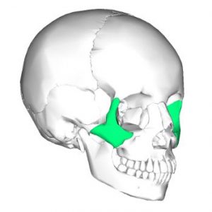
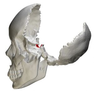
The zygomatic arch (cheekbone) is composed of the temporal bone’s zygomatic process and the zygomatic bone’s temporal process, which is joined by an oblique suture, namely zygomaticotemporal suture. Fractures of the zygoma region can occur with head trauma. In fact, the zygomatic arch is one of the most commonly fractured facial bones, typically following altercations in which the patient is punched in the face. Most zygomatic fractures are followed by a sensory abnormality, either hypoesthesia or anesthesia, in one or more branches of the infraorbital nerve. Radiographic confirmation of zygomatic arch fractures allows early stabilization with better anatomic function and cosmetic results.