Radiography of the Leg and Knee: An Overview of Anatomy and Pathology
Radiography of the lower extremity is performed to identify injuries (fractures), inflammation (osteoarthritis), weakening of bones (osteoporosis), and other conditions that may be causing pain, swelling, or inability to bear weight on the limb. Learn about the anatomy of the leg and knee joint and the pathology that can affect these parts of the body. Read about radiographic positioning of the leg and knee for the Radiologic Technologist (X-Ray Tech).
Bony Anatomy of the Lower Extremity
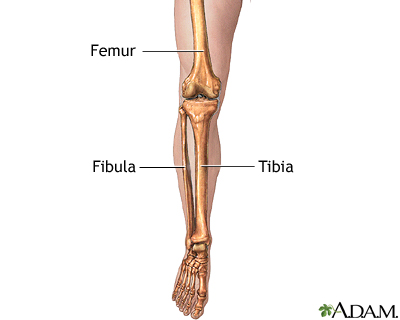
The lower limb consists of three regions, the thigh, knee joint, and leg. The thigh extends from the hip joint to the knee joint. The leg extends from the knee joint to the ankle joint. The femur is the single bone in the thigh. The tibia is the larger, weightbearing bone on the medial (inner) side of the leg. The fibula is the thinner bone on the lateral (outer) side of the leg.
Femur
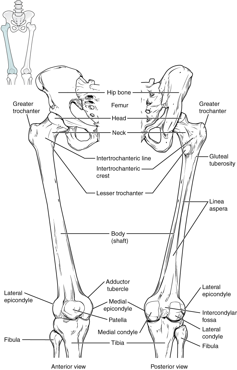
This is the strongest and longest bone in the body. It accounts for approximately a quarter of a person’s total height. At the proximal end, the femur has a rounded head which fits into the acetabulum of the hip bone to form the hip joint. The narrow region below the femoral head is the neck of the femur. This is a common area for fractures. The greater trochanter is a large bony prominence located at the base of the neck onto which many muscles attach. The lesser trochanter is a smaller prominence just below the neck. At its distal end, there are expansions on either side called the medial and lateral condyles of the femur. The epicondyles are roughened areas present on the medial and lateral condyles. The adductor tubercle is a small bump at the superior margin of the medial epicondyle. Posteriorly, there is a deep depression between the medial and lateral condyles called the intercondylar fossa.
Knee Joint

The knee joint consists of an articulation between the femur, tibia, fibula, and patella (knee cap). The articulation between the femur (thigh bone) and tibia forms the main joint. There are lateral and medial tibiofemoral compartments. The patella articulates with the femur to form the patellofemoral joint which protects the front of the knee along with the joint capsule, ligaments, menisci (thick cartilages), and bursae (fluid-filled sacs).
Tibia and Fibula
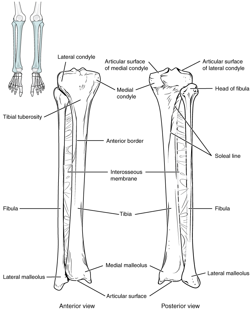
The tibia (shin bone) is the larger, inner bone in the leg. It is the second longest bone after the femur and is the main weight-bearing bone in the leg. The proximal end has expansions called the lateral and medial condyles of the tibia. There are no epicondyles like the femur. The superior surface of the medial and lateral tibial condyles is flat and smooth and articulates with the medial and lateral femoral condyles. The intercondylar eminence of the tibia is an elevated, irregular area between the condyles where supporting ligaments attach. The tibial tuberosity is a prominence near the proximal end where muscle tendons attach. The shaft of the tibia is triangular in shape. At its distal end, the tibia has a large expansion called the medial malleolus, which forms the bump on the inside of the ankle.
The fibula is the thinner bone on the lateral side of the leg. Its function is for muscle attachments. It does not bear weight. At the proximal end of the bone is a small knob-like head of the fibula which articulates with the lateral tibial condlyle to form the proximal tibiofibular joint. At its distal end, the fibula has the lateral malleolus, which forms the bump palpable on the outside of the ankle.
Common Medical Conditions Affecting the Lower Extremity
- Injuries such as muscle strain, ligament sprain, meniscus tear, hemarthrosis.
- Fractures of the femur, tibia, fibula, and patella.
- Diseases such as osteoarthritis, cysts, gout, neuroma.
- Inflammation such as bursitis, tendinitis, and synovitis.
- Deformities such as bow legs (genu varum), knock knees (genu valgum), bipartite patella.
- Dislocations of the patella or tibiofemoral joint.
- Syndromes such as iliotibial band syndrome, patellofemoral pain syndrome, Plica syndrome.
Radiography of the leg and knee plays a critical role in diagnosing and managing many of the medical conditions that affect this part of the body.
Suggested Reading
Radiographic positioning of the femur and tib fib
Radiographic positioning of the knee in AP views
Radiographic positioning of the knee in PA views
Radiographic positioning of the knee in lateral views
Radiographic positioning of the knee in patella views
RLKA
Read more about this and other subjects and get 2 Category A ARRT® CE Credits in the X-Ray CE Course “Radiography of the Leg, Knee, and Ankle”