Bone Diseases in Humans: Renal Osteodystrophy and Paget’s Disease
This article in the series on bone diseases in humans talks about renal osteodystrophy and Paget’s disease of the bone. X-ray technologists need to be familiar with these diseases as they often entail radiographic imaging. Our 2-credit ARRT®-recognized course on Radiography of the Leg, Knee, and Ankle is a great way to revise your knowledge and earn CE credits at the same time.
Renal Osteodystrophy
Patients with chronic renal disease and impaired kidney function are at risk of a complex disease known as renal osteodystrophy which affects the bones. The condition is associated with increased levels of parathyroid hormone leading to a stimulation of bone metabolism. In addition, there is a delay in the mineralization of bone due to an insufficient production of active vitamin D by the kidneys. Furthermore, some patients exhibit adynamic bone disease with a failure to form bone.
This is one of the bone diseases in humans with a complex clinical picture. A bone biopsy is often required for accurate diagnosis of renal osteodystrophy. When a patient has progressed to end-stage renal disease, the excess parathyroid hormone produces clinical manifestations such as bone cysts due to stimulation of osteoclasts.
Whereas the life expectancy of patients with end-stage renal failure can be significantly extended with dialysis, this treatment does not prevent the osteodystrophy from progressing. In fact, dialysis is associated with further abnormalities in bone metabolism, which when superimposed on the osteodystrophy lead to a high risk of fragility fractures. Kidney transplantation, on the other hand, where the patient receives a new kidney, can reverse the changes of osteodystrophy. However, immunosuppressive therapy that is necessary in transplant recipients has been linked to bone loss and increased risk of fractures. This makes renal osteodystrophy treatment one of the most complicated among bone diseases in humans.
Paget’s Disease of Bone
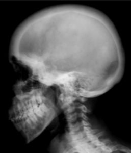
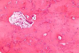
Paget’s disease of bone is a disorder of the bone remodeling process. It is a progressive condition that typically affects the skull, spine, pelvis, and legs, although any bone can be involved. Early diagnosis is critical in minimizing the impact of this disease which is one of the most crippling bone diseases in humans. Individuals with Paget’s disease of bone have overactive osteoclasts that lead to excessive loss of bone at the affected sites in the skeleton. In response to this increased bone loss, bone formation increases. However, this compensatory increase leads to the formation of new bone that is disorganized in structure. This new bone is more susceptible to fracture and deformity because not only has it expanded in size, but it also contains more blood vessels and connective tissue in the marrow.
At some sites in the body, Paget’s disease of the bone remains asymptomatic with no clinical signs. At other sites, the abnormal bone produces signs and symptoms such as deformity, bone pain, fracture, and osteoarthritis in the adjoining joints. Pagetic bone can compress nerve tissue and cause neurological complications. Very rarely, an osteosarcoma may develop and further complicate the picture.
Cause of Paget’s Disease
The pathogenesis of Paget’s disease remains unclear even though it is one of the most common bone diseases in humans, second only to osteoporosis. Although a strong genetic predisposition has been noted and Paget’s disease is known to run in families, no single gene has been identified as the culprit. Approximately 15 to 40 percent of patients with Paget’s disease have an affected family member who also has the disorder. It is therefore understood that the condition is familial and is transmitted across generations. Some studies have suggested that first-degree relatives of pagetic patients have a sevenfold higher risk of developing Paget’s disease of bone compared to people with no family history of the disorder. Nonetheless, in the majority of cases, environmental factors also play a part. Some studies have suggested that Paget’s disease of bone may be the result of a “slow” infection with the measles virus.
Paget’s Disease Symptoms
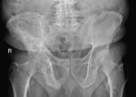
The clinical presentation of Paget’s disease of bone can vary greatly because any bone in the body can be affected by the disorder. This can make it difficult to diagnose what is one of the most common bone diseases in humans. A typical case, however, is that of a man in the sixth decade of life who presents with hip pain. Based on the patient’s age, arthritis is suspected and acetaminophen (Tylenol) and ibuprofen are prescribed. Several years later, high levels of alkaline phosphatase are noted on routine screening laboratory studies, prompting radiographs and bone scanning, which reveal pagetic changes in the patient’s pelvic bones and femur. By this time, unfortunately, there is irreversible bowing in the legs and damage to the patient’s joints.
Bisphosphonate treatment is instituted to stop disease progression, but the man must live with bone pain and walk with a limp for the rest of his life. If the diagnosis had been made earlier in the course of the disease, it could have limited the impact. Because it is known that Paget’s disease runs in families, the man’s family members are tested. A younger brother is found to have the disease, but because he receives early treatment, he never develops any deformities from the Paget’s disease.
Read more about this and other subjects and get 24 Category A ARRT® CE Credits in the X-Ray CE Course “Radiographic Bone Densitometry”DXA

Know more about x-ray continuing education here.