QRG – Imaging for Pagets Disease of Bone
Paget disease of bone is a metabolic disorder in which there is an abnormality of bone remodeling. It was first described by Sir James Paget in 1877 with the name osteitis deformans. The condition results from a focal increase in the activity of osteoclasts and osteoblasts. In advanced stages of the disease, this can spread to involve larger parts of the bone. People afflicted with Pagets disease of bone develop enlarged deformities, particularly in weightbearing regions. Susceptibility to the disease is determined by genetic factors.
How Common is Paget’s Disease of Bone?
According to The National Health and Nutrition Examination Survey (NHANES), based on surveys of radiographs of the spine, hip and pelvis, the prevalence of Paget’s disease of bone is 1.3 individuals per 100 in the 45-74 year age group. In absolute numbers, this indicates that more than 1 million Americans are affected by the condition. However, clinically diagnosed Paget’s disease is less common because, like spine fractures, Pagetic bone lesions are frequently asymptomatic. In other words, Paget’s disease of bone is present, but remains undetected.
The prevalence of Paget’s disease increases with age. Nonetheless, even in the elderly population, the incidence is less than 1 individual per 1000 annually. Interestingly, the disease affects men twice as frequently as women. Also, people of Northern European heritage are at greatest risk. Geographically, Paget’s disease of bone is found to occur most commonly in North America and Western Europe (excluding Scandinavia). There is some evidence to indicate that the prevalence of Paget’s is decreasing, although this could simply be because routine screening for the condition is not performed as frequently as it was in the past.
Stages of Paget’s Disease of Bone
- First Stage: Hot Phase: Lytic lesions
- Second Stage: Intermediate Phase: Mixed lesions
- Third Stage: Cold Phase: Dense bone
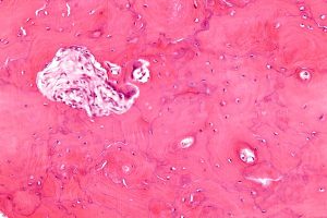
Diagnosis in the early stage is rare when osteoclastic activity predominates. The condition first affects the ends of bones and advances towards the diaphysis. However, there is one exception – the tibia – where the disease first affects the tubercle. An osteolysis zone may be visible as sharply demarcating normal bone from radiolucent fibrous tissue.
The intermediate stage is associated with mixed osteoblastic and osteoclastic activity. Trabecular coarsening and cortical thickening are indicative of new bone formation.
The cold phase consists of densification of previously laid down bone with a disorganized appearance.
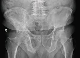
Radiographic Appearance of Paget’s Disease of Bone
In most patients, radiographic findings are diagnostic of Pagets disease of bone. Some of the radiographic features of pagetic bones include:
- Destructive bone-forming lesions
- Large well-circumscribed lytic lesions
- Cortical thickening
- Trabecular coarsening
- Multiple patches of sclerotic bone in the calvarium (cotton wool appearance)
- Enlarged and densely sclerotic vertebral bodies with prominent cortical margins
- Thickened iliopectineal line, patchy sclerosis, and lucent areas in the pelvis
- Intense uptake of radiopharmaceuticals
- Insufficiency fractures especially at high risk areas such as the femoral head and neck
- Protrusio acetabuli due to weak pagetic acetabular bone
- Multi-centric secondary sarcomas (rare complication)
However, because the appearance of Pagets disease of bone can vary greatly in different patients and at different stages of the disease, there can be other differential diagnoses such as lymphoma or metastatic deposits.
Imaging Modality of Choice for Paget’s Disease of Bone
Many of the characteristic radiographic features of Pagets disease of bone can be seen on CT and MR imaging. However, CT is usually the modality of choice as it can show complex structures in cross-sectional images, including nerve compression and sarcomatous involvement. Of course, findings seen on plain radiography can be seen in greater detail on CT. Also, MRI can demonstrate marrow signal changes that mimic the changes occurring with the disease (fatty replacement of the marrow occurs in the later stages). The disease has a mixed pattern of increase and decrease in T1 signals. Additionally, radionuclide bone scans (scintigraphy) are a sensitive imaging modality for this condition. There is marked uptake of the radiopharmaceutical in pagetic bones.
ARRT® Continuing Education Credits
Our course on bone densitometry has comprehensive information for the DXA operator as well as general radiologist. It is worth 23 ARRT® credits out of the required 24 every two years. It’s a great way to brush up your knowledge and earn most of the necessary CE credits with one easy course. Visit our online shop today. We also have courses that meet the unique requirements for radiologic technologists who perform mammography. If you’re in a hurry to get your continuing education done with, our e-course Radiography of the Upper Extremities is worth 24 Category A credits and is ideal for general radiographers. Also, keep checking back for free CE offers.
Here is details about continuing education for xray techs .