Tag: Mammography

Mammography Certification for X-Ray Techs

You can work as a mammographer when you are a radiologic technologist, but you need to be certified. The mammography certification from ARRT is intended to ensure that you have the required knowledge to work with X-rays and handle the equipment carefully. To become a mammography technologist, you need at least an associate degree as a radiologic technologist, along with an ARRT certificate. In addition, most states require candidates to have specialized training to become a mammography technologist, and earn continuing education credits.

Continue reading

Mammography Quality Standard Act Inspection Checklist

Before the Mammography Quality Standard Act was set up, there were many problems in the quality control process of mammographic equipment. Additionally, it was difficult to train mammography technologists to perform quality mammograms on patients, and inconsistencies in the system led to technicians performing suboptimal mammograms.

Therefore, to improve early breast cancer diagnosis, it was important to work on image resolution. Hence, the MQSA standards were set, and proper documentation was formed for everyone working in the mammography facility, including:

  • requirements for equipment
  • quality control
  • CE credits in both education and experience for mammography technologists, etc.

Certainly, these requirements are inspected by the MQSA inspector, and as a result, the facility is certified for providing quality mammography services to citizens. In this article, we will discuss the MQSA inspection checklist in detail to help you prepare for the evaluation day.

MQSA 1992

The Mammography Quality Standards Act (MQSA) was passed in 1992 by congress in order to achieve standard mammography quality across the nation. The decision was made to:

  • implement quality mammography services for patients
  • help in early breast cancer detection

The MQSA has been amended twice, taking into consideration the impact of setting up the requirement standards. Currently, the final amendment has detailed requirements for:

  • Employees: For example, the people who interpret mammograms, mammography techs, radio techs, physicists, etc. concerning their education, experience, and CE credits
  • Medical Audits: It is important for mammography facilities to have a system in order to follow up the obtained biopsy results
  • Quality Control and Recordkeeping: Facilities should always keep a record of their activities; certain records are required at the time of inspection

Above all, it is mandatory for facilities to get their MQSA certification in order to offer any mammography service to citizens. The facilities get certification only if they are able to comply with the MQSA requirements. If the facility fails to qualify the inspection, it has to stop providing all mammography services. Moreover, they have to regain their certificate by implementing the changes suggested by the inspector. Additionally, it is compulsory for facilities to display the valid certificate copy from the MQSA, and it must be easily visible to customers.

Who gets MQSA certified?

There is a general confusion that stays with radiologic and mammography technologists. You, as a mammography technologist or a radiologic technologist, do not get certified under the Mammography Quality Standard Act. The facility that you work in gets the certification to offer mammography services. Moreover, it is illegal to perform mammograms on people without a valid Mammography Quality Standard Act accreditation; however, it is mandatory for you as a tech to comply with the requirements set for you. You are responsible for the image resolution and comprehension of the mammograms; therefore, you will need to comply with the CE credits and the experience credits. Failure to fulfil these requirements will impact the certification for the facility.

The Inspection Checklist

The MQSA inspector informs the facility well in advance about the inspection date and time. The facility needs to prepare in such a way as to still be able to provide services to patients. The inspector will need approximately 20 minutes in each mammography unit to evaluate the mammogram. The following is a checklist for you to crosscheck your preparations for the inspection day.

Pre-Inspection requirements

  • Keep records of every activity that you perform
  • Document all the information
  • Keep ample space ready for the inspector to view the documents undisrupted

Reports

  • All other inspection reports that are conducted by the state within the last and the current MQSA inspection period
  • Safety Survey Reports of the equipment
  • Record retention policy
  • State registration documents upholding the present name, contact, and owner details
  • Availability of all the required protective shields for employees and equipment

Medical Records

  • Display all records; the inspector will select any 5 of his choice
  • Keep a medical record log that will help track positive results
  • Have letter samples to depict the use of easy language in result demonstration
  • Documentation to ensure that the physicians review all the reposts

Quality Control

  • EQUIP reports
  • Ensure that all the monitors and QC manuals are updated to the latest versions
  • Put forward all the QC manuals and QC tests of all the monitors (on-site and off-site) used since the last inspections
  • Ensure that the information is the same in the QC forms as it is in the one filed in the monitor software
  • Provide Medical Equipment Evaluation reports about all the units, new and old
  • Provide reports about the evaluation of the mamo unit after the replacement of any part.

Plan of Action Manual

  • Display the technique chart
  • Provide detailed information on the responsibilities associated with the role of every employee
  • Keep records of service and operator manuals of all mamo units
  • Keep physicist reports
  • Keep meeting minutes of the quality assurance meetings held at the facility to improve the service quality

Credential Requirements for Employees

On-site and Off-site Medical Physicists

Initial Qualifications

  • Degree certificate, 20 hours per semester in physics, 20 contact hours in mammography surveys
  • Should have survey 1 facility and 10 units or an FDA approval letter
  • Unexpired State Approval / License
  • 2-D and 3-D initial training – 8 CEU’s (only applicable if your facility uses the same)

Continuing Education / Experience Requirements

  • 15 CEUs in 3 years
  • Should have maintained a record of surveying at least 2 facilities and 6 units in 24 months

Radiologic Technologist

Initial Qualifications

  • Copy of the 40 hours of training and the ARRT R card
  • Have supervised 25 mammograms
  • 8 CE units of the 2-D and 3-D training (if used in your facility)

Continuing Education / Experience

Interpreting Physicians

Initial Qualifications

  • Copy of a valid MD license and ABR
  • 3 months of training and 60 CMEs (if qualified after 4/28/99)
  • 40 CMEs and 2 months of training
  • Should have supervised 240 readings within a period of 6 months
  • 8 CEUs in 2-D and 3-D initial training (only applicable if your facility uses them)

Continuing Education / Experience

  • Should have given readings of at least 960 mammograms in 2 years
  • Should have earned 15 Continuing Education Units in 3 years
  • Additionally, the CEUs must be in category 1

Conclusion

MQSA inspection is an annual affair, which aims to maintain a standard quality of mammography services around the country. The evaluation is complete only with the exit interview that the inspector gives. The inspector might inform you about the deficiencies during the exit interview or you may receive an email containing the result of the inspection.

OPTO-ACOUSTIC BREAST IMAGING

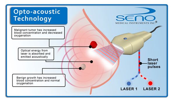
Opto acoustic breast imaging is a new generation diagnostic tool currently in the clinical trial stage for diagnosis of malignant or benign breast lumps. According to a European journal of breast cancer around 4 million new cases of breast cancer are detected every year with almost 80 percent of them turning out to be benign on laboratory testing. It is difficult for radiologists to rule out malignancy at the imaging stage. The cost of conducting surgical or core needle biopsy for every suspicious lump outweighs the results especially in women coming from struggling economies. A low cost diagnostic tool such as opto acoustic imaging can help in the timely detection of breast cancer in the early stages and also help prevent and control the increasing mortality due to this disease in developing as well as developed countries.

Continue reading

Scheduling a Mammogram

By: Stacey Nester

Effective communication with patients begins at the very moment a mammogram appointment is scheduled. It is essential to instill confidence in patients to not only achieve the highest quality images but also to ensure they have a positive and reassuring experience at your facility.

It is vital that personnel responsible for scheduling are thoroughly trained to handle communications with mammogram patients in a sensitive and caring manner. This approach is crucial for providing support and ensuring a positive interaction from the very first contact.

Continue reading

Establishing Patient Rapport During Mammograms

Understanding the Emotional Landscape of Mammography

Due to the deeply personal and sensitive nature of mammography, it is challenging to anticipate the various emotions or personal circumstances a woman may be facing during her appointment. Each individual’s experience can significantly differ based on a myriad of factors unique to her situation.

Factors Influencing Emotional Response

Several factors can shape a woman’s emotional state during a mammography appointment:

  • Family History of Breast Cancer: Women with a family history of breast cancer may feel heightened anxiety or fear.
  • Previous Health Scares: Past health issues can increase apprehension.
  • Physical Discomfort: Concern about the physical discomfort associated with the procedure.
  • Potential Outcomes: Anxiety about the results of the mammogram.

“Each individual’s experience can significantly differ based on a myriad of factors unique to her situation.”

Cultural and Personal Beliefs

Cultural and personal beliefs about health and medical procedures can also influence a woman’s emotional state:

  • Embarrassment or Discomfort: The intimate nature of the examination can cause feelings of embarrassment or discomfort.
  • Financial Concerns: Worry about the potential financial implications of the procedure and follow-up care.

Experience with Mammograms

Previous experience with mammograms can shape expectations and comfort levels:

  • First-Time Patients: May feel more anxious due to unfamiliarity with the process.
  • Returning Patients: May have different expectations and levels of comfort based on past experiences.

For more information on mammography and tips on managing the emotional aspects of the procedure, visit our Radiologic Technology Resources page.

Explore continuing education opportunities in radiography at CE4RT.

Approaching Mammography with Sensitivity and Empathy

Healthcare providers must be aware of the varied emotional landscapes women may bring to their mammography appointments. Each experience is unique, and approaching each appointment with sensitivity and empathy is crucial.

Effective Communication

Effective communication can help alleviate anxiety and discomfort. Providers should:

  • Provide Clear Information: Explain the procedure thoroughly.
  • Create a Supportive Environment: Ensure the patient feels comfortable and supported.

“Understanding that each patient’s experience is unique and offering personalized care can make a significant difference.”

Personalized Care

Recognizing the unique emotional landscape of each patient is essential. Personalized care includes:

  • Approaching each patient with openness and sensitivity.
  • Establishing a positive rapport to ease anxiety, especially given the intimate nature of the exam.

Addressing Emotional Concerns

Technologists should be mindful of the range of emotions patients may experience, including:

  • Fear of Potential Results: Address concerns about the outcomes with compassion.
  • Discomfort with the Procedure: Acknowledge and mitigate physical discomfort.

Enhancing the Mammography Experience Through Understanding and Compassion

Understanding and addressing patients’ feelings can significantly enhance their mammography experience. Patients may feel vulnerable or anxious due to a family history of breast cancer or personal health scares. Technologists can play a key role in alleviating these feelings.

Active Listening and Clear Communication

Technologists can help by:

  • Listening Actively: Pay attention to patients’ concerns and fears.
  • Providing Thorough Explanations: Clearly explain the procedure to demystify the process.
  • Reassuring Patients: Set realistic expectations to reduce apprehension.

“Transparency in communication can demystify the process and reduce patient apprehension.”

Creating a Supportive Environment

Support goes beyond technical expertise and includes:

  • Showing Empathy and Patience: Allow time for patients to express their concerns.
  • Maintaining a Calm and Friendly Demeanor: Offer words of encouragement and ensure privacy and comfort.
  • Responding with Compassion: Address concerns with understanding and care.

The Impact on Diagnostic Outcomes

Prioritizing these aspects of patient care helps alleviate some of the emotional burden associated with mammography. This approach improves the overall experience for the patient and contributes to more accurate and effective diagnostic outcomes, as a relaxed and cooperative patient is more likely to comply fully with necessary positioning and instructions during the exam.

Creating a Supportive Environment During Mammography

Acknowledging and validating the patient’s feelings is key to fostering a supportive and comfortable environment. This is especially important during mammography, which can be both physically and emotionally demanding. The physical discomfort of breast compression, combined with anxiety about potential findings, can make the experience particularly challenging for many women.

The Importance of Understanding Emotions

Recognizing and addressing patient emotions helps technologists play a crucial role in making patients feel more secure and cared for. This starts with:

  • Active Listening: Hear the patient’s concerns and respond empathetically.
  • Validating Feelings: Acknowledge that the procedure can be uncomfortable and that their feelings are normal.

“Simple acts of validation can go a long way in reducing anxiety.”

Providing Clear Explanations

Offering clear, concise explanations about each step of the procedure can help patients feel more in control. For example:

  • Explain the Procedure: Detail why compression is necessary for obtaining clear images.
  • Highlight the Benefits: Help patients understand how compression contributes to accurate diagnosis.

Impact on Patient Experience

By recognizing and addressing these emotions, technologists can create a more positive experience for patients. This approach not only reduces anxiety but also encourages patient cooperation, leading to more accurate and effective diagnostic outcomes.

Enhancing Patient Comfort During Mammography

Technologists play a crucial role in improving patient comfort and reducing anxiety during mammography. Small adjustments and a compassionate approach can make a significant difference.

Making Adjustments Based on Patient Feedback

Listening to and acting on patient feedback can greatly enhance comfort. Here are some strategies:

  • Adjust Positioning: Modify the patient’s position to reduce discomfort.
  • Take Breaks: Allow breaks if the patient feels overwhelmed.

“Providing reassurance and maintaining a calm, compassionate demeanor throughout the appointment can further alleviate stress.”

Providing Reassurance and Maintaining Calm

Maintaining a supportive atmosphere involves:

  • Offering Reassurance: Continuously reassure the patient throughout the procedure.
  • Compassionate Demeanor: Stay calm and compassionate to help alleviate patient stress.

The Impact on Imaging Results

By acknowledging and validating the patient’s feelings, technologists not only improve the emotional experience but also encourage better cooperation. This can lead to:

  • Improved Cooperation: Patients are more likely to follow instructions accurately.
  • More Accurate Imaging: Reduced anxiety can result in clearer and more accurate images.

Creating an environment where patients feel understood and supported helps ensure that mammography is as positive and stress-free as possible.
Continue reading