Tag: imaging

OPTO-ACOUSTIC BREAST IMAGING

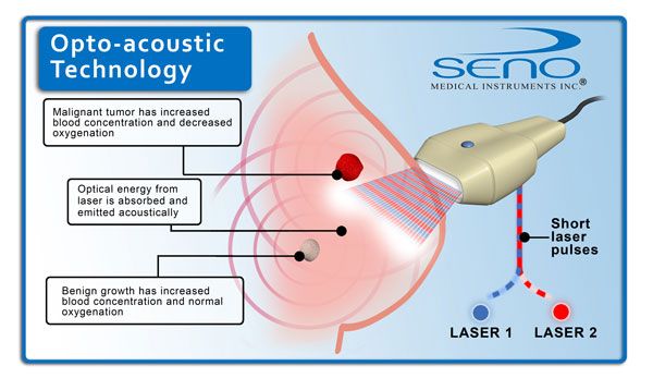
Opto acoustic breast imaging is a new generation diagnostic tool currently in the clinical trial stage for diagnosis of malignant or benign breast lumps. According to a European journal of breast cancer around 4 million new cases of breast cancer are detected every year with almost 80 percent of them turning out to be benign on laboratory testing. It is difficult for radiologists to rule out malignancy at the imaging stage. The cost of conducting surgical or core needle biopsy for every suspicious lump outweighs the results especially in women coming from struggling economies. A low cost diagnostic tool such as opto acoustic imaging can help in the timely detection of breast cancer in the early stages and also help prevent and control the increasing mortality due to this disease in developing as well as developed countries.
Continue reading

TJC Diagnostic Imaging Standards Revision 2014

By: CE4RT


In early 2014, The Joint Commission (TJC) announced changes to its standards for accredited hospitals, critical access hospitals, and ambulatory health care organizations that provide diagnostic imaging services. TJC has stated changes will be effective some time in 2015 with additional requirements to be phased in by 2015. The standards changes announced by TJC, however, in most cases will not require any change in action by imaging centers, since most of the requirements are already being followed by imaging centers and radiology departments.

Continue reading

An X-Ray Tech’s Guide to Radiography in Obese Patients

By: CE4RT

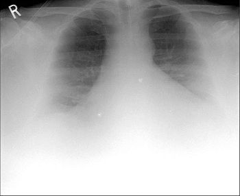
Obese patients are at risk for increased radiation exposure due to the increase in radiation needed to penetrate tissue, the risk for repeat exams due to difficult positioning and technique, an incompatibility with existing equipment, and because obese patients suffer from health conditions which require radiologic procedures more than the average public.


A 57 year old man who weighed 490 lb (223 kg). Chest radiograph shows limited diagnostic quality image with poor X-ray penetration and poor visualization of lung bases.

Continue reading