X-ray Equipment Safety Features: Essential Information for Radiologic Technologists
Radiologic technologists use a number of methods to protect against ionizing radiation. This includes the use of shielding and following the ALARA principle. However, an important component of radiation safety are the X-ray equipment safety features in the imaging devices that X-ray techs operate.
The FDA is responsible for the pre-market safety evaluation of digital radiography equipment. The goal is to ensure that the equipment delivers optimum radiation dose and image quality. The goal is also to ensure that the equipment has appropriate dose-reduction features and automatic notification of high dose rates. Manufacturers of X-ray equipment must meet certain design requirements to ensure radiation safety. For imported equipment, the supplier must ensure that the equipment meets the regulatory requirements of the United States in addition to regulations in the country of origin.
Safety Features
Some of the X-ray equipment safety features include:
- Beam shutter (prevents the primary beam from passing when closed)
- Failsafe (turns off X-ray generator and closes the beam shutter)
- Beam stop (absorbs the primary beam)
- Interlock (shuts down the beam if any switch in a series is open)
- Warning lights (indicate X-ray beam is on)
- Warning signs (caution that radiation-producing equipment is present)
The X-ray device is labeled with the make, model, serial number, and maximum operating parameters along with the manufacturer’s name and address. The position of the focal spot is marked on the tube housing. The tube housing and support are immobilized and locked in position. In addition, the following radiation safety or dose-reducing features are present in digital radiography equipment:
X-ray Tube Housing
In addition to enclosing the components and dissipating heat, the X-ray tube housing restricts X-ray emission to the useful primary beam. Therefore, it is an important X-ray equipment safety feature. The protective housing is constructed to limit leakage to no more than 100 mR/hour at a distance of 1 meter from the source when operated at maximum voltage and current.
Beam-Limiting Devices
A dose-limiting feature of radiographic equipment is a manual or automatic beam-limiting device (collimator) that is attached to the X-ray tube. A collimator narrows the primary beam and confines the irradiated field to the area being studied to prevent unnecessary irradiation of tissues not being examined. When the beam is focused on the correct anatomy, it reduces both patient dose and scatter.
Positive Beam Limitation
If the radiographic beam is larger than the image receptor, some tissue is irradiated but not recorded. If the radiographic beam is smaller than the image receptor, part of the area of interest may not be recorded. Positive beam limitation is an X-ray equipment safety feature in digital radiographic equipment that ensures the radiation field and image receptor are of the same size. This ensures all the irradiated anatomy of diagnostic interest is recorded on the image receptor.
Filtration
Low-energy rays in the X-ray spectrum contribute to patient dose but do not reach the image receptor and do not contribute to image formation. Therefore, digital imaging equipment has inherent and added filters to absorb low-energy rays and improve the quality of the X-ray beam, i.e., make it more penetrating (this is known as beam hardening).
Control Console
The control console is located behind a protective barrier and indicates the exposure parameters and source-to-image receptor distance. The console has the main on-off switch for flow of current to the tube. The console has a provision to terminate exposure immediately and after a pre-determined time. This makes it one of the most important X-ray equipment safety features.
Automatic Exposure Control
In addition to manual selection of exposure factors, such as tube current, tube voltage, and exposure time, digital radiography equipment has automatic exposure control (AEC) for consistent reproducible exposure in patients of different sizes. In in terms of radiation protection, X-ray exposure is always initiated manually, but the AEC can be programmed to terminate exposure after a pre-determined dose.
Anti-Scatter Grid
Digital imaging devices have a detachable anti-scatter grid that is used when imaging parts of the body that are more than 10 cm thick. The use of a grid improves image quality but is associated with an increase in patient dose.
Examination Table
The examination table in radiographic equipment is made of carbon fiber material which is strong, stable, and radiolucent. A uniformly thick, radiolucent table is desirable because when the table absorbs minimal radiation, less radiation needs to be produced to form the image. This reduces the patient’s dose. An under-table configuration, where the X-ray tube is positioned under the patient, produces less intense scatter, making it a useful X-ray equipment safety feature.
Emergency Stop Button
All X-ray equipment has an emergency stop button which, when pressed, immediately cuts electrical power to the generator. This conspicuous red button is used in emergency or accident situations. All radiology technologists must be familiar with the position of the emergency stop button and the procedure for immediate shutdown. It is important to use this button only in emergencies, and not routinely as an off switch. If the emergency stop button is accidently activated, the operator should seek the help of the support engineer before attempting to restart the generator.
Modality-Specific Safety Features
In addition to the above-mentioned generic safety features in digital radiographic equipment, there are certain modality-specific X-ray equipment safety features. Digital radiography equipment is provided with sufficiently long electrical cables to allow safe operation and location of the console. Digital detectors are periodically calibrated per the manufacturer’s recommendations. Interventional radiology equipment and C-arm equipment features a meter that displays cumulative air kerma, dose-area product, number of cine runs, total fluoroscopy time, and exposure parameters.
Some machines may have built-in X-ray equipment safety features such as ceiling-suspended leaded-glass shielding flaps. Fluoroscopic procedures are associated with some of the largest radiation doses in diagnostic radiology. Fluoroscopy equipment is therefore provided with an indicator of cumulative time to keep the dose below the deterministic threshold. Fluoroscopy equipment also has a foot-operated pressure switch and a visual indicator on the console that lights up when the beam is on. Digital fluoroscopy equipment includes radiation safety features such as pulsed mode and last image hold for dose reduction.
CT and Mammography
In CT equipment, the control console indicates section thickness and pitch factor in addition to filtration and exposure parameters. Exposure control (beam initiation and continuation) is only possible from the control console. An emergency stop switch is located near the gantry and is an important X-ray equipment safety feature. It can be pressed to immediately terminate emission of X-rays and movement of the equipment. Parameters such as CTDI and DLP are displayed on the console to monitor and record patient dose. In equipment with more than one focal spot, the focal spot that is selected is clearly indicated on the console.
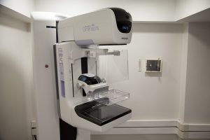
Mammography equipment includes hands-free controls for power-driven compression paddles that release compression immediately when exposure is terminated. The viewing box for mammography equipment has higher luminescence. PACS and other digital radiological equipment is interconnected to exchange information per international and national standards such that there is no loss or unintentional alteration of patient information. These are all equipment features that enhance safety in digital radiography.
X-ray Equipment Safety Laws and Regulations
A number of policies and procedures are applicable to radiation facilities to ensure the safe use of digital radiographic equipment. These policies include mandatory registration of facilities that utilize radiation-emitting machines. Registration of new X-ray equipment, annual renewal of registration for existing equipment, and registration of any changes made to the equipment (for example, replacement of the X-ray tube) are also necessary, and this is the responsibility of the radiation safety officer at a healthcare facility. The facility must notify the relevant authority when equipment is moved, leased, transferred, stolen, or lost. State and local inspectors perform periodic equipment surveys. X-ray facilities are also subject to regulatory inspections. The RSO must make all appropriate records available at the time of an inspection.
Authorized Users
The medical director of a healthcare facility approves the responsible users who are authorized to operate X-ray equipment. Some physicians and technologists may only have restricted privileges (for example, limited to performing non-radiating examinations such as ultrasound examinations). All personnel who operate X-ray equipment must have documented training in X-ray equipment safety features and the safe operation of the devices they will be using. The X-ray equipment should be used in accordance with the manufacturer’s guidelines and for the intended purpose only. During imaging, the patient must remain in clear view and the operator should be able to communicate with the patient and any attendants. All personnel present in the room should be notified by the operator before switching on the X-ray device. X-ray machines are never to be left unattended when they are producing radiation.
Shielding and Room Design
X-ray room shielding is approved by the state or local radiation control agency at new facilities or following remodeling at existing healthcare organizations. At the time of inspection, records related to X-ray shielding, such as measurement of radiation behind shielded walls, must be presented. Different states have varying requirements regarding warning signs, interlocks, restricted access, and lead equivalent thickness of shielding material. Medical physicists oversee X-ray equipment safety features. The necessary qualifications for medical physicists vary from state to state. Service providers who install, repair, and test radiographic equipment must be vetted by the RSO and approved through a state-specific process. It is the RSO’s responsibility to maintain all equipment records according to the regulations of the state radiation safety control agency.
The duration for which calibration, modification, maintenance, and equipment survey records need to be preserved also varies from state to state. Facilities where research using radiation machines is performed are subject to approval by institutional review boards. Local and state regulations may also require radiation facilities to monitor patient skin dose for lengthy fluoroscopic procedures, particularly if the dose is likely to exceed 2-6 Gy, in which case it may be mandatory to inform the patient and the radiation safety agency.
X-ray Continuing Education
We offer a range of courses with category A credits that will fulfil your ARRT® CE requirements and are accepted by NMTCB, ARDMS, SDMS, every US state and territory, Canadian province, and all other Radiologic Technologist, Nuclear Medicine, and Ultrasound Technologist registries in North America for both full and limited permit technologists, guaranteed. You can choose a course based on the number and type of credits you need. Still got questions? Read our FAQ section or contact us for all your X-ray CE queries.
Visit here to get more information about radiography continuing education.