DXA Scan Errors: Do You Know These Common Sources?
Bone density testing is an important tool in the management of osteoporosis. Several different radiological modalities are available to measure bone density. Although each one has its own advantages and disadvantages, DXA is the most widely used. However, there are certain DXA scan errors that DXA operators and general radiographers should be aware of.
BMD Measurement with Different Modalities
The BMD results obtained from different modalities cannot be compared. Even when the technology is the same, cross-calibration is necessary to compare the results from different devices. Therefore, the skill of the radiologic technologist plays a large role in limiting DXA scan errors. Precision errors are inherent to BMD testing.
BMD Testing and Precision
The term precision refers to the reproducibility of the BMD result. It is calculated by scanning 15 patients thrice in one day or 30 patients twice on the same day. Repositioning of the patient must be done following each scan. The precision calculation is used to calculate the least significant change (LSC). Therefore, it is important for every DXA facility to perform precision assessment. Facilities must also perform comparison of BMD measurements between different radiologic technologists to reduce DXA scan errors. Moreover, the manufacturer’s recommendations for quality control and maintenance should be adhered to. Finally, errors in BMD testing creep in because the education and training of technologists for bone densitometry varies widely.
Sources of DXA Scan Errors
The sources of DXA scan errors can be categorized based on technique, bone disease, and artifacts. Errors due to technique include patient positioning, movement, and ROI placement. Those due to bone diseases include fractures, sclerotic or lytic lesions, and degenerative changes. Mistakes due to artifacts include foreign bodies such as calcifications, surgical material, and contrast media. Broadly speaking, there are four possible sources of DXA scan errors: the instrument, the patient, the technologist, and the report.
Instrument Errors
Operators should be aware that differences exist between different machines and between the same machine installed at different centers. In addition, algorithms and reference databases vary from manufacturer to manufacturer. It is therefore not appropriate to directly compare the results of the current report to a prior report obtained on a different machine or at a different facility. DXA scan error due to equipment include hardware/software problems such as tissue typing errors or image errors (detector streaks). Mechanical problems include phantom and QA issues, atypical sounds from the device, and software error messages. In addition, the environment of the instrument, such as temperature, humidity, power outages, and power surges, can lead to mechanical problems.
Patient Error
The technologist performing the bone densitometry study should be aware that the patient’s height and weight, i.e., body mass index, have an effect on BMD. This is because the manufacturer’s algorithms are based on a normal BMI and are not valid for extremely underweight or extremely overweight individuals. Failure to keep this in mind can result in DXA scan errors. In addition, the presence of metal objects on the patient can produce artifacts. The patient should also be questioned regarding recent exposure to contrast or nuclear medicine investigations.
Technologist Errors
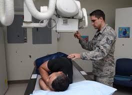
Positioning of the patient by the technologist is critical for accurate results. Failure to position correctly can result in DXA scan errors. In the hip, for example, rotation of the femur affects the area of bone being scanned. Studies have found that internally rotating the hip to various degrees other than the standard 15-20 degrees produces significant changes in BMD. For spine scans, the selection of the region of interest is critical and can be a source of error even with experienced technologists. Deformities and osteophytes in the vertebrae can make it difficult to define vertebral levels and lead to inconsistent findings. It is also worth noting that errors can occur due to different operators in the same center performing patient positioning and analysis differently.
Report Errors
A DXA report contains a large amount of information. Radiologic technologists should be aware that there is potential for misunderstanding here. Being vigilant can prevent these types of DXA scan errors. Technologists should be able to identify and understand all the items on a DXA report. It is important for DXA operators to record the reference data being used to ensure correct interpretation by the radiologist.
Common Mistakes in DXA Scanning
Listed below are some the most common DXA scan errors and mistakes in BMD testing. These can have potential adverse effects on the patient’s clinical management.
Indications
Common mistakes in this category include not performing BMD testing in a high-risk patient. For example, an asymptomatic 67-year-old woman is at risk on account of her age and gender and should undergo BMD testing even in the absence of any symptoms. Other common DXA scan errors are to perform bone densitometry in a patient whose clinical management is unlikely to change based on the results, for example, a healthy 35-year-old woman.
Quality Control
Failing to follow the device manufacturer’s instructions for system maintenance and failure to conduct phantom measurements are common QC DXA scan errors. If phantom scan results are not reviewed or phantom scanning is not done following recalibration, errors can creep in. Significant changes in calibration must be identified and corrected. Precision assessment and calculation of least significant change (LSC) must be performed. Quantitative comparison of BMD tests is only possible if LSC is known.
Scan Acquisition
Incorrect patient positioning and selecting the wrong scan mode are common DXA scan errors. Other errors include measuring BMD at an invalid skeletal site and not removing artifacts from the scan area. Entering the wrong patient demographic information is yet another mistake that can occur during scan acquisition.
For example, if the spine is not parallel to the long axis of the DXA table or the hip is not properly internally rotated, the BMD measurement will be inaccurate. If BMD is measured in a patient who has had total hip replacement, this result is not usable. A patient’s underwire bra within the scan field is an artifact error. Entering the wrong age/gender is another common mistake. Choosing the wrong scan mode can lead to inaccurate results. The correct scan mode should be chosen according to the size of the patient (a longer scan time is required for larger patients with denser bones).
Analysis
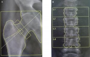
Review and correction of default bone edge, ROI identification, and incorrect labeling of vertebral bodies are common DXA scan errors. Radiologic technologists should make use of bony landmarks such as the iliac crest and lower ribs to correctly label the vertebrae. Review and manual correction of the default bone edge is necessary. This is because the computer sometimes includes a large osteophyte in the scanned area of the spine.
Interpretation
The improper application of the WHO diagnostic criteria for osteoporosis is a possible interpretation error. Stating there is bone loss based on a single BMD test is another mistake. Fracture risk may sometimes be incorrectly calculated. These are all interpretation DXA scan errors.
Common Artifacts in DXA Scanning
Some of the common artifacts encountered clinically that can lead to DXA scan errors are listed below:
- Patients with limb amputations may have missing bone and soft tissue in the region of interest.
- Patients who have undergone internal fixation of the spine.
- Bracelets, watches, anklets.
- Battery pack of external hearing aid in pocket. Internal hearing aids are typically too small to affect the scan.
- Penile implant.
- Breast implants.
- Total or partial hip or knee replacement.
- Plaster of Paris or fiberglass cast.
- Movement artifact, which may require rescanning.
- Obesity which can cause excessive noise and affect areal BMD precision.
Checklist for DXA Technologists
Operators can use the following checklist before performing a scan to help avoid DXA scan errors:
- Can the patient tolerate lying on their back for 10-20 minutes?
- Is the patient’s weight within the acceptable limit for the DXA table?
- Has the patient had any imaging procedure with contrast in the past two weeks?
- Is the patient premenopausal, and if so, is there a possibility of pregnancy?
- Has the patient taken calcium tablets in the past 24 hours?
- Is the patient appropriately dressed in comfortable, loose-fitting clothes?
- Are there any metallic components such as zippers, rivets, or underwire in the patient’s clothes?
- Has the patient brought the results of prior bone densitometry tests with them?
- For body composition studies, is the patient fasting overnight (12 hours) for consistency?
- Do I have a printout of the previous image/baseline scan to ensure consistency in positioning and scan parameters?
DXA Continuing Education
We have a range of online courses for DXA CE available for purchase. These e-courses are recognized by the ARRT® and other registries and are good to complete the required X-ray CE credits. Our DXA CE online course and test is an easy and convenient way to earn 23 CEUs of the required 24 every two years. Choose one of our other courses to complete your requirements.
Read more about this and other subjects and get 24 Category A ARRT® CE Credits in the X-Ray CE Course “Radiographic Bone Densitometry”DXA
Here is details about california fluoroscopy ce credits.