Ionizing Radiation Video Series
We discovered this video series and thought it might be of interest to this community. There is no CE credit for this series, but it is useful for reference.
Radiologic Technologist Blog
We discovered this video series and thought it might be of interest to this community. There is no CE credit for this series, but it is useful for reference.
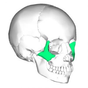
The zygomatic arch (cheekbone) is composed of the temporal bone’s zygomatic process and the zygomatic bone’s temporal process, which is joined by an oblique suture, namely zygomaticotemporal suture. Fractures of the zygoma region can occur with head trauma. In fact, the zygomatic arch is one of the most commonly fractured facial bones, typically following altercations in which the patient is punched in the face. Most zygomatic fractures are followed by a sensory abnormality, either hypoesthesia or anesthesia, in one or more branches of the infraorbital nerve. Radiographic confirmation of zygomatic arch fractures allows early stabilization with better anatomic function and cosmetic results.

Quantitative computed tomography (QCT) is a medical technique that measures bone mineral density (BMD) using a standard X-ray Computed Tomography (CT) scanner with a calibration standard to convert Hounsfield Units (HU) of the CT image to bone mineral density values. Quantitative CT scans are primarily used to evaluate bone mineral density at the lumbar spine and hip.

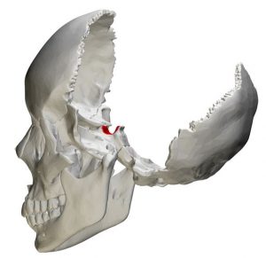
The sella turcica (also called the hypophyseal fossa or pituitary fossa) is a midline saddle-shaped depression in the sphenoid bone that is lined by the dura mater. Although it is a relatively small area, it is an extremely valuable piece of real estate in the brain because it forms the bony seat for the pituitary gland which it houses and partially encloses. One of the main reasons for imaging the sella turcica is that it is a window to the pituitary, a pea-sized gland that is often called the master endocrine gland because of the major role it plays in regulating vital body functions. Sellar components are easily demonstrated by several radiographic planes and angles. Radiology techs need to be aware of the anatomy of this region as well as correct radiographic angles and patient positioning techniques to demonstrate the sella turcica and surrounding structures accurately.
Getting an order for mastoids is rare these days. But it still happens occasionally, and when it does don’t be ashamed to pull out your Merril’s. Tissue thickness, superimposing shadows, and awkward patient positioning make the mastoid process a difficult body part to radiograph.
If you really want to make your mark as a radiologic technologist, a wide variety of customized Left and Right X-Ray markers and supplies are available online.
If you recently purchased a course but are having trouble seeing it below, it’s most likely a caching issue. Try clicking the button below to refresh the page. If that doesn’t work, follow the manual instructions below based on your operating system:
Windows/Linux:
Hold the Ctrl key and press the F5 key.
Or, hold down Ctrl and ⇧ Shift and then press R.
Mac:
Hold down the ⇧ Shift and click the Reload button.
Or, hold down ⌘ Cmd and ⇧ Shift and then press R.