Diagnostic Reference Levels and Other Metrics
Any individual who undergoes a medical imaging examination is subject to the benefits as well as the risks of the examination. Each individual is unique, with a unique body habitus, health status, and medical needs. Because of the wide variety and complexity of medical conditions, dose limits cannot be set for medical exposures. However, the average radiation exposure associated with a diagnostic or interventional procedure can be used to assess whether the dose received by the patient is within acceptable range. In other words, estimated exposure levels for different examinations help to highlight any substantial variation from the expected value. Certain metrics such as diagnostic reference levels make this possible.
Our course on ALARA in Digital Radiography has further information on radiation safety and is worth 4 ARRT® credits that count towards CEU for radiology technologists.
Diagnostic Reference Levels
Exam-specific and region-specific exposure levels are known as diagnostic reference levels (DRLs) or diagnostic acceptable reference levels (DARL-ings). DRLs are representative of the acceptable exposure for an average-sized patient during normal practice in the local area.
DRLs are advisory levels indicated by a numerical value. They serve as tools for dose audit. For digital radiographic examinations, the recommended DRL parameters are kerma area product and entrance surface AK. They are not regulatory, but rather voluntary guidelines. These levels are issued by the ACR and AAPM based on NEXT survey data. National and local DRLs should be reviewed periodically when new digital equipment is introduced. DRLs need to be refined for clinical image quality and the body weight/size of the patient population typically imaged at a facility.
In routine clinical practice, it is expected that the typical dose for the majority of examinations will be less than the established reference level. DRLs thus ensure patient radiation safety by serving as a benchmark figure against which doses from diagnostic procedures performed at a facility can be compared. Diagnostic reference levels help avoid excessively high radiation doses to patients. DRLs do not apply to each individual patient or each individual examination performed at a facility. However, if a DRL is repeatedly being exceeded for a certain type of exam, it calls for a review of the technique followed at the facility.
Action Limits
Action limits are not the same as diagnostic reference levels. Action limits are exposure levels established by a healthcare organization to trigger an evaluation and determine the cause for an unusually high exposure to an individual patient or member of staff.
Dose Constraints
Dose constraints are used to restrict procedure optimization if the overall exposure will remain within the dose limit. They are used to optimize occupational exposure and facility design. It should be noted that dose constraints are different from diagnostic reference levels.
Dose Index Registry
Dose information is automatically extracted from DICOM image headers and stored in this central registry of doses established by the ACR. The registry can be queried by individuals or facilities to obtain their dose indices for specific procedures and compare them against local, regional, national, and international values. The radiation safety committee at a healthcare organization can use the registry to periodically evaluate the radiation associated with a specific imaging study. A dose index that is too low may be indicative of inadequate image quality. A dose index that is too high may be indicative of unnecessary use of excessive radiation.
Negligible Individual Dose
The NCRP introduced the idea of negligible individual dose. This is the effective dose associated with average excess risk of fatal health effects per year that can be attributed to radiation. In other words, NID is the cutoff level for radiation exposure, below which the effective dose is of negligible risk. Therefore, efforts to reduce the exposure below the NID are not warranted. The NID concept is used to develop radiation protection measures for X-ray systems used in scanning. Americans are exposed to roughly 6.2 millisieverts of radiation per year from natural sources and medical exposures.
The NCRP recommends keeping additional radiation exposure from non-medical, non-natural sources to under 1 millisievert per year. The negligible individual dose is defined as less than 0.01 millisieverts. NID is an important metric that can be used in conjunction with diagnostic reference levels to ensure radiation safety without wasting resources.
Detective Quantum Efficiency
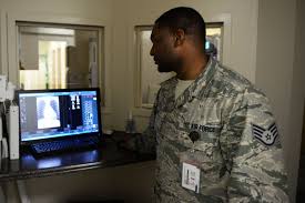
Detective quantum efficiency (DQE) quantifies the combined effects of the signal associated with the image contrast and noise performance of an imaging system, generally expressed as a spatial frequency function. This value is basically used to describe imaging detectors in the fields of medical radiography and optical imaging.
In medical radiography, the detective quantum efficiency describes the effectiveness of an X-ray imaging system when it comes to producing an image with high signal-to-noise ratio (SNR) relative to an ideal detector. Sometimes, it is considered as an alternative measure of radiation dose efficiency of a detector since the required radiation exposure to a patient (as well as the biological risk of that particular radiation exposure) is reduced, as the DQE increases for the same image SNR and exposure conditions. It features a combination of very low noise and superior contrast performance, which allows some digital X-ray systems to provide such relevant improvements in terms of the detectability of low-contrast objects. This characteristic is best measured by a single parameter, known as DQE. In fact, according to a medical physics expert, the DQE has become the benchmark when comparing the technologies of an existing and an emerging X-ray detector. Unlike diagnostic reference levels which are patient-based, DQE is equipment-based.
The DQE method mainly affects one’s ability to view small, low-contrast objects. In most imaging situations, it is more important to detect small objects rather than using a limiting spatial resolution (LST) to determine the size of an object one can visualize. Although digital systems usually have high LSR, they can’t take advantage of the resolution if they have low DQE, which averts the detection of very small objects.
Detective Quantum Efficiency in Film/Screen vs Digital Imaging
There was research that compared film/screen to digital imaging. It showed that a digital system having high DQE can enhance one’s ability to perceive small, low contrast objects despite the fact that the digital system may have a relatively lower LSR (limited spatial resolution) than films.
Another advantage of digital x-ray technology is its ability to reduce radiation dose. High DQE should heavily contribute to this equation. In contrast to film screen imaging, high DQE digital detectors have the potential to convey significant improvements in object detectability at an equivalent dose or to allow object detectability, as compared to film, at a lower dose.
Moreover, high DQE provides the necessary foundation for advanced digital applications, such as low-dose fluoroscopy, dual-energy imaging, and tomosynthesis. When DQE is combined with fast acquisition, rapid readout capability, and advanced image-processing algorithms, it becomes a key essential process in making these applications more clinically practical in the coming years. This can make it an important metric to be used in conjunction with diagnostic reference levels.
Area Surveys
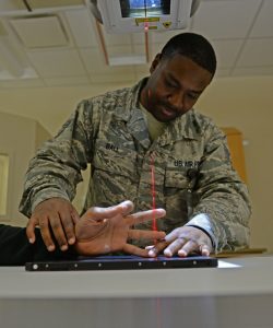
To comply with radiation safety regulations, healthcare facilities that use X-ray equipment and radioactive materials are required to develop, implement, and document precautionary procedures known as area surveys. Such surveys assess the prevailing radiological conditions and help to identify potential hazards. Whereas diagnostic reference levels are patient-centric, area surveys are location-centric. Surveys consist of a combination of calculations and measurements. For example, ambient radiation exposure rate is measured with radiation detection instruments. Surface wipe tests are performed to detect contamination or residual radioactivity. Licensees are required to maintain records of surveys. The requirement for retaining these records may differ from state to state but is typically three years after the record is generated.