Radiographic Positioning of the Lumbar Spine
This article discusses radiographic positioning of the lumbar spine for the Radiologic Technologist (X-Ray Tech).
- Lumbar Spine AP or PA
- Lumbar Spine PA Ferguson Method
- Lumbar Spine Lateral
- Lumbar Spine Lateral Supine
- Lumbar Spine AP Oblique
- Sacrum AP
- Lumbosacral Junction Lateral
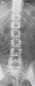
Radiologists consider a lumbar spine radiographic film of good quality when it demonstrates the lower ribs, lumbar vertebral bodies, transverse processes, pedicles, spinous processes, sacrum, and sacroiliac joints. On lateral projections, the intervertebral disc spaces and intervertebral foramen as well as the superior and inferior articular processes should be visible along with the vertebral bodies and spinous processes.
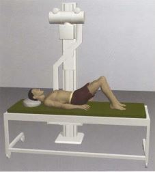
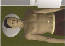
Lumbar Spine AP or PA
Purpose and Structures Shown A basic view of the lumbar spine.
Position of patient Supine or prone. Injured patients should NOT be turned over. In trauma patients, a lumbar spine X-ray is done in the AP or PA position with minimal movement of the patient.
Position of part The patient’s knees are bent to ensure the back is flat on the table. The patient should be asked to stop breathing when the exposure is taken.

Video Credit : Vien Chunggia
Lumbar Spine PA Ferguson Method
Purpose and Structures Shown An alternate view of the lumbar spine in PA projection to protect radiosensitive organs from exposure. This is a scoliosis series that is used to distinguish a primary deforming curve from a compensatory curve.
Position of patient Standing or seated in front a vertical grid. The IR should be adjusted to include about 1 inch of the iliac crests. The midsagittal plane of the body should be aligned to the midline of the grid. The arms hang by the side of the body in the standing position. The elbows are flexed and the hands rest on the lap in the seated position. A second radiograph is taken with the hip or foot elevated with a block or sandbag under the foot or buttock. The Ferguson method requires the patient to expend some effort to maintain this position without support.
Position of part The patient should be asked to suspend respiration during exposure. The gonads are shielded. The image shows the thoracic and lumbar vertebrae in PA projection with the vertebral column in the center of the radiograph.
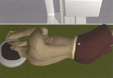
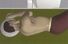
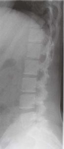
Lumbar Spine Lateral
Purpose and Structures Shown A basic view of the lumbar spine.
Position of patient Lying on the left or right side (lateral recumbent) with the knees and hips flexed for comfort. The elbows are flexed and the arms are at a right angle to the body. The knees are superimposed. The midcoronal plane is aligned to the midline of the grid. In patient’s with suspected fracture, lumbar spine supine with horizontal beam should be used instead.
Position of part The gonads are shielded. The patient should be asked to breathe in, breathe out, and then hold the breath during the exposure. The image demonstrates the lower thoracic to coccyx area with the lumbar vertebral bodies, intervertebral disc spaces, spinous processes, and lumbosacral junction visualized.
Video Credit : Vien Chunggia
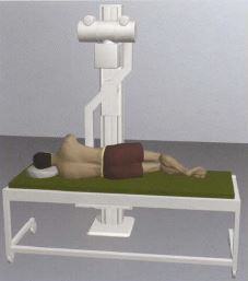
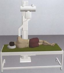
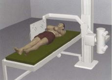
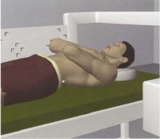
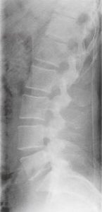
Lumbar Spine Lateral Supine
Purpose and Structures Shown An additional view of the lumbar spine for patients with injuries.
Position of patient Supine with a horizontal beam. The midsagittal plane is centered to the middle of the grid. The hips and shoulders are in the same plane horizontally. The elbows are flexed and the hands are placed on the upper chest to remove the forearms from the exposure field. The patient may be asked to flex the knees and hips to bring the back in firm contact with the table and reduce lumbar lordosis.
Position of part The gonads are shielded. The patient should be asked to breathe OUT and hold respiration during exposure. The image demonstrates the lumbar vertebral bodies, intervertebral disc spaces, transverse and spinous processes, pedicles, and laminae. The sacroiliac joints are seen on either side at equal distance from the vertebral column. The vertebrae are symmetric with the spinous processes centered on the bodies. In trauma patients, a larger field may be used to enable visualization of additional abdominal organs such as the kidneys, liver, spleen, and psoas muscle.
Lumbar Spine AP Oblique
Purpose and Structures Shown An oblique projection that is usually performed after the AP projection to demonstrate the articular processes and/or lumbosacral processes.
Position of patient Supine and turned 45 degrees towards the affected side. The long axis of the body should be parallel to the long axis of the table. The spine is centered to the midline of the grid. The lumbar spine is approximately 2 inches medial to the elevated anterior superior iliac spine in the oblique position. The arms are in a comfortable position. The patient should be asked to hold the breath during exposure.
Position of part A support may be placed under the elevated parts of the body (shoulder, hip, knee). The gonads are shielded. The degree of body rotation should be 45 degrees to demonstrate the articular processes and 30 degrees to demonstrate the lumbosacral processes. The image shows an oblique projection of the area from the lower thoracic vertebrae to the sacrum. The articular processes on the side closest to the IR are visualized. Zygapophyseal joints are open and uniformly seen through the vertebral bodies. The other side is imaged for comparison.
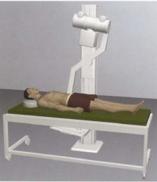
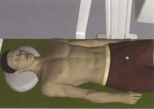
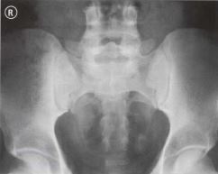
Sacrum AP
Purpose and Structures Shown A basic view of the lumbosacral junction and sacroiliac joints.
Position of patient Supine with a vertical beam angled at 15 degrees. This view is NOT used in children. Gas and fecal matter in the bowel can interfere with images of the sacrum and coccyx. A bowel preparation (by physician’s order) may be needed. The bladder should be emptied prior to the examination.
Position of part The tube should be angled at 15 degrees. Centering should be done 3 cm above the pubic symphysis. The patient can breathe normally during exposure. The gonads are shielded in men. Female gonads cannot be shielded for this projection. The image clearly demonstrates the sacrum free of superimposition.
Video Credit : Vien Chunggia
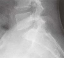
Lumbosacral Junction Lateral
Purpose and Structures Shown A basic view of the lumbosacral junction. This view is NOT used in children.
Position of patient Lying on the left or right side. The patient should be asked to bend the knees to stabilize the body. A pad should be placed under the waist for support. If possible, the hips should be fully extended.
Position of part Centering should be done 3 cm below the iliac crest. The knees are exactly superimposed. The patient should be asked to suspend respiration during exposure. The gonads are shielded. The image demonstrates the lumbosacral joint in the center of the image. The entire L5 and upper sacrum should be visualized.