Radiographic Positioning of the Thoracic Spine
This article discusses radiographic positioning of the thoracic spine for the Radiologic Technologist (X-Ray Tech).
- Thoracic Spine AP
- Thoracic Spine Lateral
- Thoracic Spine AP or PA Oblique Projection Upright
- Thoracic Spine AP or PA Oblique Projection Recumbent
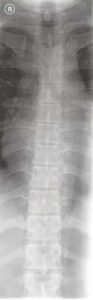
In addition to the thoracic vertebral bodies, spinous processes, and transverse processes, a good quality thoracic spine AP projection shows the left ventricle, gastric bubble, right/left hemidiaphragm, posterior ribs, and right/left clavicle. The lateral projection shows the left and right hemidiaphragm, vertebral bodies, intervertebral disc spaces, and posterior ribs.
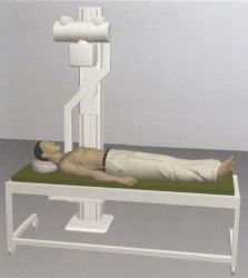
Thoracic Spine AP
Purpose and Structures Shown A basic view of the thoracic spine showing all 12 thoracic vertebral bodies, intervertebral disc spaces, transverse processes, and costovertebral joints. Two radiographs may be necessary for the upper and lower thoracic regions.
Position of patient Supine. The arms are placed by the side of the body and the shoulders are flat on the table. The head should rest directly on the table or on a thin support to avoid exaggerating the thoracic kyphosis.
Position of part The gonads are shielded. The patient is asked to stop breathing when the exposure is taken. The vertebral column is aligned to the middle of the image. Ribs, shoulders, diaphragm, and lungs are visible.
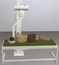
Thoracic Spine Lateral
Purpose and Structures Shown A basic view of the thoracic spine.
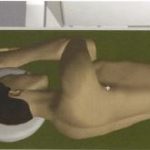
Position of patient Lying on the left or right side (lateral recumbent position). The left lateral position is preferred because placing the heart closer to the IR minimizes overlap.
Position of part The patient is positioned with knees bent to stabilize the body. The long axis of the spine is horizontal. The gonads are shielded. The patient may breathe normally when the exposure is being taken.

Video Credit : RadPositioning
Thoracic Spine AP or PA Oblique Projection Upright
Purpose and Structures Shown An oblique projection of the thoracic spine to demonstrate the zygapophyseal articulations.
Position of patient Standing or sitting erect in a lateral position in front of a vertical grid. The body is rotated 20 degrees posterior or anterior (AP or PA oblique, respectively). The vertebral column is centered to the midline of the grid. The shoulder adjacent to the grid rests firmly against it. The arm adjacent to the grid is positioned as follows: For PA oblique, the elbow is flexed and the arm rests on the hip; for AP oblique, the arm is extended forward to prevent superimposition of the humerus on the upper thoracic vertebrae. The outer arm is positioned as follows: For PA oblique, the patient is asked to grasp the grid; for AP oblique, the outer hand is placed on the hip.
Position of part The long axis of the vertebral column is parallel to the IR. The gonads are shielded. During the exposure, the patient is asked to hold the breath at the end of expiration. All 12 thoracic vertebrae should be clearly demonstrated along with the zygapophyseal joints.
Thoracic Spine AP or PA Oblique Projection Recumbent
Purpose and Structures Shown An oblique projection of the thoracic spine to demonstrate the zygapophyseal joints.
Position of patient Lateral recumbent. The head is elevated on a pillow. The patient’s knees and hips are flexed for comfort. For the PA oblique projection, anterior rotation is obtained by extending the upper arm forward and placing the hand on the table and placing the lower arm behind the back. For the AP oblique projection, posterior rotation is obtained by placing the upper arm posteriorly with support and positioning the lower arm at a right angle to the body with the elbow flexed. The body is rotated by 20 degrees anteriorly or posteriorly.
Position of part The gonads are shielded. During the exposure, the patient is asked to hold the breath at the end of expiration. All 12 thoracic vertebrae should be clearly demonstrated along with the zygapophyseal joints.
Visit here to know more about radiographic positioning and related anatomy.