Tag: radiography

Radiography of the Sella Turcica

By: CE4RT

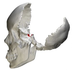
Location of the sella turcica in the skull
Sella Turcica Location in the Skull

The sella turcica (also called the hypophyseal fossa or pituitary fossa) is a midline saddle-shaped depression in the sphenoid bone that is lined by the dura mater. Although it is a relatively small area, it is an extremely valuable piece of real estate in the brain because it forms the bony seat for the pituitary gland which it houses and partially encloses. One of the main reasons for imaging the sella turcica is that it is a window to the pituitary, a pea-sized gland that is often called the master endocrine gland because of the major role it plays in regulating vital body functions. Sellar components are easily demonstrated by several radiographic planes and angles. Radiology techs need to be aware of the anatomy of this region as well as correct radiographic angles and patient positioning techniques to demonstrate the sella turcica and surrounding structures accurately.

Continue reading

Nuclear Medicine CQR and Structured Education

By: CE4RT


To ensure quality healthcare, the American Registry of Radiologic Technologists® (ARRT®) has implemented a system of periodic evaluation consisting of Structured Education and Continuing Qualification Requirements (CQR). Radiologic Technologists certified before January 1, 2011, are not affected by these new requirements. Technologists who were certificate on or after 1/1/2011 receive a time-limited 10-year license. Once the 10 years are up, certification can be renewed for another 10 years by completing the CQR process. In addition, compliance with the ARRT® rules and regulations includes an annual renewal process and biennial continuing education requirements. R.T.®s in Nuclear Medicine require specific CE credits related to their certification. The ARRT® calls this structured education.

Continue reading

Radiography and Pregnancy

By: CE4RT

All radiographers are trained to understand the risks of radiation exposure to the fetus of a pregnant patient and the importance of identifying pregnant patients. This allows them to inform the physician about the risks, ensuring that all necessary precautions are taken during imaging procedures.

If you are a radiologic technologist who becomes pregnant, it’s natural to be concerned about the potential harmful effects of radiation exposure to your unborn child. The question often arises: Do X-ray techs need special precautions during pregnancy? While the evidence strongly indicates that radiographers can safely continue their duties without risking fetal health, provided they adhere strictly to safety policies and guidelines, it is still crucial for every radiographer to be well-informed about the potential effects of radiation exposure during pregnancy.

Pregnant Radiographers and Radiation Workers

Pregnant radiographers and radiation workers face heightened health risks due to the sensitivity of rapidly developing embryos and fetuses to radiation. As a result, their maximum allowable radiation exposure is strictly limited to 0.5 rem (or 5 millisieverts) over the entire gestation period. This safety threshold is just 10% of the typical annual dose limit for radiation workers, which is set at 5 rem (or 50 millisieverts).

Continue reading

5 keys for Digital Radiography

By: CE4RT

Computed Radiography (CR) and Direct Radiography (DR) are now the standard imaging technologies in many hospitals and imaging centers. These digital systems mark a significant evolution in the field of medical imaging. X-ray technicians must grasp several critical concepts to effectively utilize these technologies. Familiarity with the functionalities, advantages, and specific operational aspects of both CR and DR systems is essential for maximizing image quality and ensuring patient safety. Each system has unique applications and demands specific skills and knowledge for effective management. Learn more about the capabilities and applications of digital radiography systems here.

Continue reading