Quick Refresher for X-ray Techs: Cardiac Screening Guidelines
Screening Tests for Coronary Artery Disease
Coronary artery disease is the most common type of heart disease. It occurs when plaque builds up in the arteries that supply the heart muscle. The American Heart Association has outlined key screening tests to monitor cardiovascular health. Cardiac screening guidelines include the following tests to detect CAD in people with no signs and symptoms:
- Serum lipids
- Blood glucose
- C-reactive protein (indicator of swelling or inflammation)
- Blood pressure
Until the 1980s, only 1 in 3 Americans knew their cholesterol number. In the following decades, healthcare systems, public health agencies, and voluntary organizations nationwide made coordinated efforts to target screening for chronic medical conditions such as heart disease. A program called the National Cholesterol Education Program was implemented in the mid-1980s. The objectives was to keep healthcare providers updated on current evidence-based cardiac screening guidelines. Also, the program encouraged increased awareness in the general public about the adverse effects of high cholesterol. By the mid-1990s, about 75 percent of Americans “knew their number,” i.e., cholesterol level.
Several decades later, the incidence of heart disease has continued to climb. More sophisticated cardiac screening guidelines have been developed to detect heart disease before symptoms begin. These guidelines and procedures are important because the initial symptom of heart disease is often a serious medical event such as a heart attack. Heart disease is the leading cause of death in the United States. Early detection, lifestyle changes, and patient education can reduce the health consequences of this medical condition. Radiologic technologists play a key role in the heart health of the American public.
Screening Tests for Coronary Artery Disease
Coronary artery disease is the most common type of heart disease. It occurs when plaque builds up in the arteries that supply the heart muscle. The American Heart Association has outlined key screening tests to monitor cardiovascular health. Cardiac screening guidelines include the following tests to detect CAD in people with no signs and symptoms:
- Serum lipids
- Blood glucose
- C-reactive protein (indicator of swelling or inflammation)
- Blood pressure
Additional Screening Tests and Imaging Procedures
Based on the presence of risk factors (age, gender, family history, smoking, diabetes, obesity, hypertension, high cholesterol) and the results of the above-listed screening tests, patients are often sent to the radiology department for additional testing and imaging procedures, including:
- EKG to measure the electrical activity of the heart.
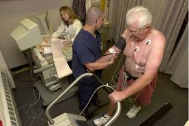
- Cardiac stress test or exercise EKG to determine if there is adequate blood flow to the heart when it is stressed.
- Echocardiography to visualize a moving picture of the heart with ultrasound.
- Cardiac CT for calcium scoring to measure the amount of calcium (an indicator of the presence of plaque) in coronary arteries.
- Coronary CT angiography with contrast dye to see the exact location and severity of plaque buildup in the coronary arteries.
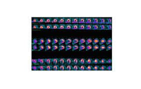
- Myocardial perfusion imaging or nuclear stress test with radioactive material to see the effect of stress on blood flow and heart muscle.
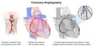
- Coronary catheter angiography to see any narrowing or blockages in the coronary arteries.
Cardiac CT for Calcium ScoringThis test is included in cardiac screening guidelines because it is noninvasive and indicates whether a patient is at increased risk of a heart attack. No injection of contrast is required. The test is painless and takes relatively little time. CT examination exposes the patient to some radiation but no radiation remains in the patient’s body after the test is completed. Cardiac CT for calcium scoring documents the presence or absence of plaque in the coronary arteries. This information is used to guide treatment.
For the procedure, the patient is positioned supine on the CT examination table. Pillows and straps may be used to maintain the correct position and help the patient remain still during the CT scan. An EKG machine is put in place. The patient is asked to raise their arms above the head and hold their breath at specific times as the CT scanner obtains multiple images.
The CT technologist should ask all women of childbearing age whether they are or could be pregnant. CT scans are not recommended during pregnancy in general due to the potential risk to the baby. However, a CT scan may be performed on a pregnant woman in special circumstances when it is unavoidable. Patients should be informed that radiation exposure increases the lifetime risk of cancer slightly. Nonetheless, the benefits outweigh the risks and all precautions are taken to minimize radiation exposure as much as possible.
Coronary CT Angiography (CTA)
CTA is a noninvasive test included in cardiac screening guidelines. It is an alternative to cardiac catheterization with coronary angiogram which is an invasive test and associated with a higher number of complications. CTA can detect or exclude plaque buildup and blockages in the coronary arteries. Also, this test can visualize soft tissues and bones in addition to blood vessels. This helps in detecting other causes of symptoms, for example, aortic injury or pulmonary embolism (blood clot in the lungs). Additional advantages include that CT examinations are quick and painless. Also, CT is less sensitive to patient movement than MRI and can be performed in patients with implanted medical devices. There is radiation exposure during the examination, but no radiation remains in the patient’s body afterward.
At the start of the procedure, an IV line is inserted into the patient’s arm. A medication is given to slow down the heart rate. Nitroglycerine is given by spray or sublingual (under the tongue) to widen the coronary arteries. This helps obtain better images. The EKG machine is set up to record electrical activity. Contrast dye is given through the IV line. Patients are asked to raise their arms above the head and hold their breath at specific times during the examination. The CT scanner is used to obtain multiple images.
Risks of CTA
Potential risks of coronary CT angiography include worsening of kidney function due to the contrast material in people with kidney disease. Also, there is a risk of contrast extravasation (leaking of the dye under the skin). CT technologists should advise patients to immediately inform them if they experience any pain in the arm during the IV contrast injection. As noted above, CT scanning is not recommended in pregnant women. According to the American College of Radiology, it is safe for mothers to breastfeed their babies, although contrast manufacturers say breastfeeding should be avoided for 24-48 hours after dye administration. There is also a risk of rare, but serious allergic reaction to contrast materials. CT technologists should obtain a careful history from patients recommended to undergo procedures per cardiac screening guidelines. A previous history of reaction to CT contrast may require premedication with steroids.
Myocardial Perfusion Imaging or Nuclear Stress Test
This procedure is included in cardiac screening guidelines because it provides unique information about the structure and function of the heart muscle. Oftentimes, this information is not obtainable from other imaging procedures and it helps in making a diagnosis and/or determining the appropriate treatment.
The examination begins by giving the patient a small amount of radioactive material called the radiotracer. After waiting for 20-40 minutes, the patient is positioned on the table and an IV line is inserted. The patient is asked to raise one or both arms above the head during the imaging at specific times. Afterward, the heart is monitored while the patient walks on a treadmill or pedals a stationary bike. At the time of peak blood flow to the heart, another injection of radiotracer is given, followed by 20-40 minutes of rest. In patients who are unable to exercise, a drug to increase blood flow to the heart is given. Finally, a second series of images is obtained.
Patients with coronary artery disease may experience some discomfort or chest pain during the exercise or when the medication to increase blood flow is given. In such cases, the patient should be carefully monitored and a medication for chest pain may be given. Potential risks include the extremely rare occurrence of allergic reaction to the radiopharmaceutical agent. Any pain and redness due to radiotracer injection typically resolves quickly. The technologist should question women of childbearing age about pregnancy and breastfeeding. Radiation exposure from this test increases the lifetime risk of cancer, but this risk is small and the benefits outweigh the risks.
Coronary Catheter Angiography
This test is included in cardiac screening guidelines because it provides a clear, detailed picture of the cardiac vasculature to a degree that is not available with any other noninvasive examination. It is often performed before procedures such as percutaneous interventions. Unlike CTA, it is possible to combine diagnosis and treatment with coronary catheter angiography. For example, a severe narrowing may be detected in a coronary artery, an angioplasty performed, and a stent deployed, all in a single procedure. The radiation exposure associated with this test does not usually have any side effects.
At the start of the examination, an IV line is inserted into a vein in the patient’s arm or hand. The groin area where the catheter will be inserted is cleaned and shaved and numbed with local anesthetic. The cardiologist makes a small incision in the groin area and guides the catheter to the heart. Contrast material is injected through the IV line and multiple X-ray images are obtained.
Potential risks of coronary catheter angiography include:
- Allergy to contrast dye
- Contrast extravasation
- X-ray exposure in pregnant women
- Contrast exposure of babies in breastfeeding mothers
- Formation of clot at the tip of the catheter, blocking the artery, and requiring an operation to open the vessel
- Worsening kidney function due to contrast administration in people with kidney disease
- Puncture of the artery by the catheter leading to internal bleeding
- Catheter tip separates material from the artery’s inner lining, causing blockage downstream
- Increased risk of cancer from radiation exposure
X-ray Continuing Education
We hope you found the information in this article on cardiac screening guidelines and procedures useful. We offer a range of ARRT®-recognized continuing education courses online. Browse our website for specialized e-courses for DXA operators, mammographers, and fluoroscopists. Each course is worth between 1 and 24 CEUs, giving you the flexibility to choose the type and number of credits you need.
Here is details about ce credits for x-ray techs and xray continuing education.