Patient Radiation Dose Measurement in Fluoroscopy and CT Examinations
Radiation dose represents the energy deposited per unit mass of tissue. It is typically measured in Gray (Joules/kg). This deposition of energy may cause damage the tissues, and therefore, patient radiation dose must be measured and monitored. Patients are exposed to some of the largest doses of radiation during fluoroscopically-guided procedures and computed tomography examinations. Radiologic technologists play a key role in limiting this dose, based on the ALARA principle, by using the correct imaging techniques.
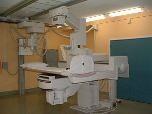
Measuring Radiation Exposure from Fluoroscopic Procedures
During fluoroscopically-guided examinations, patients are exposed to sufficiently large radiation doses and it is important to measure the radiation dose and document it in the medical record. It is also wise to monitor the patient post-procedure for possible deterministic effects (cutaneous radiation injury). The primary problem in measuring patient radiation dose from fluoroscopically-guided procedures is that dose administration is not uniform throughout the body. In addition to total fluoroscopy time, certain metrics such as peak skin dose and kerma area product have been developed to quantify radiation exposure. Some of these metrics are good for estimating stochastic risk (kerma area product), while others are good indicators of risk for deterministic effects (peak skin dose). You can learn more about patient radiation dose and limiting exposure with our course ALARA in Fluoroscopy.
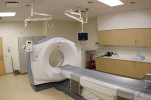
Measuring Radiation Dose from CT Procedures
During CT examinations, the main concern is stochastic effects since doses that can cause deterministic effects are generally not reached during these types of studies. To determine the stochastic effect, we must identify which tissues were exposed and how much radiation they got. This information is contained in a metric called Effective Dose, the unit of which is Sieverts (Sv). This measurement of patient radiation dose takes into account the fact that a dose of 1 Gy to the brain is not the same as a dose of 1 Gy to the foot because these tissues have different radiation sensitivities. Because CT involves shooting X-rays at different angles into the body, we need to measure the dose through metrics such as CT dose index. Let’s take a more detailed look at some of the metrics used to measure radiation dose.
Peak Skin Dose
This measures the dose, including backscatter, absorbed by a specific point on the surface of the patient’s body (i.e., the skin). It is expressed in milligray (mGy). PSD cannot be measured directly and must be estimated geometrically from the signal to object distance and air kerma. Peak skin dose indicates the maximum dose at one point on the surface of the patient’s body in the radiation field. PSD is affected by several factors, including the shape and size of the field, the gantry position and angle, and the size and positioning of the patient. Peak skin dose is important because it is used to identify patients who are at potential risk of severe deterministic effects (local reactions) from radiation exposure and may require followup and monitoring for cutaneous radiation injuries. The NCRP, the ICRP, and various other regulatory bodies have emphasized that estimating peak skin dose is essential for the management of acute radiation injury. Any skin dose in excess of 15 Gray is considered a sentinel event.
Surface Absorbed Dose
This metric is used to assess the risk of cutaneous radiation injury such as skin erythema. It is the radiation energy deposited in one unit of mass at the skin entrance surface. Surface absorbed dose (SAD) is expressed in milligrays. This measure of patient radiation dose is particularly important in interventional cardiology procedures.
Total Air Kerma
Kerma is an expansion of the phrase “kinetic energy released in mass.” Total air kerma indicates the amount of radiation energy (in joules) absorbed by a unit mass of air (in kg) at one point in relation to the source of the beam. The unit of air kerma is Joules/kg or Gray, which is also the unit for absorbed dose. Exposure can be converted to kerma with the help of conversion coefficients. The reference point for air kerma (AK) is calculated according to the inverse square law and is located at the entrance port on the patient at a fixed distance from the center of the beam.
Therefore, cumulative AK measures the actual radiation output from the source. If the reference point where air kerma is measured is the point where the beam strikes the patient’s soft tissue, then the absorbed dose and air kerma are theoretically the same. However, there is considerable variation between cumulative air kerma and peak skin dose. This is because any movement leads to a spread of the beam across the body surface and an alteration in peak skin dose. On the other hand, cumulative air kerma is unaffected by movement as it measures radiation at a single point in air. Cumulative air kerma is NOT a good measure of radiation exposure. This is because, firstly, the established reference point does not always correspond to the actual location of the beam on the patient’s body surface. Secondly, cumulative air kerma does not take into account scatter, tissue absorption, and beam attenuation.
Surface Integral Exposure
Radiation exposure and air kerma provide information about the concentration of radiation delivered to a specific point. These quantities do not give any information about the total amount of radiation absorbed by the body. The surface integral exposure (SIE) is the exposure in mR multiplied by the exposed area in cm2 when exposure is uniform over an area of the body. When exposure is not uniform, the SIE is calculated by adding up the individual exposure area products for the whole body. SIE is important because the stochastic risk of radiation-induced cancer is related to the total radiation that enters the body and not the surface concentration of the beam striking the body. As noted, the surface concentration of the beam at a specific area of the body is a better indicator of the risk of cutaneous radiation injury.
Dose Area Product
This is also known as kerma area product (KAP). It is measured in Gray-cm2 and is a surrogate measure of the energy deposited by a beam of X-rays. DAP is similar in concept to surface integral exposure but is expressed in different units. DAP remains constant irrespective of the distance between the patient and the source of X-ray beam. Dose area product estimates the total radiation energy that a patient receives during an imaging procedure. DAP is used to estimate stochastic risk (i.e., the risk of radiation-induced cancer) and is a practical method of monitoring the total amount of radiation delivered to a patient. The disadvantage of DAP is that a large area exposed to a low dose and a small area exposed to a high dose may have the same DAP. Like radiation exposure, air kerma, and SIE, the DAP can be measured with an ionization chamber. Many angiography and fluoroscopy units have an ionization chamber attached to the collimator to directly measure DAP.
Computed Tomography Dose Index
This is a specific quantity used to indicate the radiation dose associated with a CT imaging procedure. During CT imaging, the X-ray beam rotates around the patient and multiple contiguous scans are obtained with uniform exposure. This means that the absorbed dose from a single central slice is a good indicator of the dose delivered to the tissue. Measurement protocols for CTDI take into account the exposure from the primary beam as well as from scatter radiation. CTDI is calculated with the help of a phantom and TLD dosimeter or ionization chamber. This quantity is called a dose index because it is not a precise measurement of dose but rather an approximate measure or estimation. The weighted CTDI is a measurement that takes into account the dose variance across a CT image. Weighted CTDI is calculated with TLD dosimeters placed both centrally and peripherally in the phantom.
Computed Tomography Dose Length Product
This is a quantity that is accepted all over the world. It specifies the total radiation delivered to a patient during a CT imaging procedure. It is obtained by multiplying the CTDI value by the length of anatomy scanned. The units for DLP are Gy-cm or rad-cm. It is a useful metric to compare the radiation dose associated with different CT procedures. DLP is displayed on many CT scanners to aid the technologist in achieving dose optimization.
Multiple Scan Average Dose
This is the average dose from each slice at the mid portion of a multi-slice scan. MSAD is an accurate measure of radiation dose at the central portion of a series of CT scans. The dose is highest at the center. Therefore, at either end of a series, MSAD overestimates the patient’s radiation dose. MSAD takes into account the fact that overlapping scans are associated with a higher dose and gaps between CT slices lower the patient’s radiation dose. The process to calculate MSAD is laborious and requires multiple scan acquisitions. CTDI is a simpler and more practical method to estimate radiation output from computed tomography systems. When the slice thickness and bed index (image spacing) are equal, then MSAD and CTDI are equal. It is noteworthy that neither CTDI or MSAD give an estimate of the patient’s total dose, but only the average radiation dose in a scan volume.
ARRT® CE for X-ray Techs
We have a range of online courses for X-ray CE. All our e-courses are recognized for ARRT® category A credits. Choose a course to conveniently and easily complete your biannual continuing education requirements.
Read more about this and other subjects and get 12 Category A ARRT® CE Credits in the X-Ray CE Course “ALARA and Radiation Protection”UIRP
Here is more information about radiography continuing education.