Description
Get 24 Bone Densitometry CE Credits now. Guaranteed**.
This course satisfies both Genral Radiography CE Credits and DXA CE Credit requirements in all 50 states and Canada. It offers with ARRT®*
Category A credits as well as CQR for DXA (aka DEXA). This course meets ARRT®* continuing education requirements for Bone Densitometry CQR Credits (Continuing Qualification Requirements).
Course Objectives
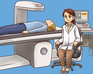
Students of this course will be instructed to read the E book Radiographic Bone Densitometry. The book contains up to date and comprehensive information regarding osteoporosis, bone densitometry, radiation protection, and procedural tactics. Upon completion of the reading material, there is a post test that needs to be completed in order to receive a certificate of completion.
The student, having completed the course, will have the latest information on radiographic bone densitometry and radiation protection for professionals, as well as tools and techniques for minimizing radiation dose for patients and staff, and quality control.
Course Outline
Section 1 – Patient Care
Osteoporosis – Definition of Osteoporosis, Primary Osteoporosis, Secondary Osteoporosis, WHO Diagnostic Criteria of Osteoporosis
Bone Anatomy – Bone Marrow, Periosteum, Endosteum, Cortical Bone, Cancellous (Trabecular or Spongy) Bone
Bone Physiology – Structural Support and Protection, Storage of Essential Minerals, Osteoblasts, Osteocytes, Osteoclasts, Bone Growth, Bone Remodeling Cycle, Factors Affecting Bone Remodeling,
Impact of Nutrition and Exercise on Bone Health – Role of Calcium and Vitamin D in Bone Health, Role of Other Nutrients in Bone Health, Impact of Exercise and Physical Activity on Bone Health
Osteoporosis and Bone Health Risk Factors – Factors Affecting Bone Mass, Changes in the Skeleton at Different Stages of Life, Impact of Weight on Bone Health, Reproductive Factors Affecting Bone Health, Hysterectomy and Oophorectomy, Effect of Smoking, Alcohol, and Heavy Metals on Bone Health, Diseases and Osteoporosis Risk, Medications That Impact Osteoporosis Risk
Prevention and Treatment of Osteoporosis – Lifestyle Factors, Fall Prevention, Drug Therapies, Hormone Therapy, Risks versus Benefits of Postmenopausal Hormone Therapy, Selective Estrogen Receptor Modulators, Calcitonin, Combination Therapy with Antiresorptive Drugs, Combination Therapy with Anabolic Agents and Antiresorptive Drugs, New and Future Therapies for Osteoporosis, Laboratory Tests and Biochemical Markers, Study Design for Osteoporosis Treatments, Antiresorptives, Drugs that Promote Bone Formation, Osteoporotic Fractures, Incidence of Fractures, Treatment of Osteoporotic Fractures, Future Research on Population-Based Interventions
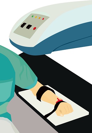
Imaging Modalities for Bone Densitometry – Digital X-Ray Radiogrammetry (DXR), Quantitative Ultrasound, Quantitative Computed Tomography, Photon Absorptiometry, Single-Energy X-Ray Absorptiometry (SXA), Dual-Energy X-Ray Absorptiometry (DXA aka DEXA), Next Generation Risk Assessment Tools, Radiography, Multi Detector Computed Tomography (MDCT), High resolution Peripheral QCT, Magnetic Resonance Imaging, DXA Operator Training and Certification, Bone Mass Measurement Act, ICD Codes, CPT Codes
Patient Preparation for DXA – Patient Preparation for DXA, Patient Instructions, Explanation of Procedure, Motion and Breathing Requirements, Number and Duration of Scans, Patient History, Medical history, Contraindications, Clinical Indications and Guidelines, Patient Factors, Special Needs, Fall Prevention and Mobility Assistance, Mental Impairment or Disorientation, Removal of Artifact Producing Clothing, Anatomy, Pathology, Body Habitus, Nonremovable Artifacts, Pediatric and Adolescent DXA Scanning, Procedural Considerations with Pediatric DXA, DXA Measurement in Newborn Babies, Indications for DXA in Pediatric and Adolescent Patients
Section 2 – Safety
Fundamental Principles of Radiation Safety – ALARA, Basic Methods of Protection, Time, Distance, Shielding
Biological Effects of Radiation – Deterministic Effects, Skin Injury from Radiation, Cataracts, Sterility, Acute Radiation Syndrome, Teratogenesis, Stochastic Effects, Carcinogenesis 183, Radiosensitive Tissues / Organs
Units of Measurement – Absorbed Dose, Equivalent Dose, Effective Dose, Skin Dose, Levels of Radiation in DXA
General Radiation Protection Issues – Scatter Radiation, Occupational Protection, Room Design and Scanner-Operator Distance, Personnel Monitoring, Exposure Records
Patient Radiation Protection – Comparison Levels of Radiation, DXA Dose, Peripheral or Axial, Natural Background Radiation, Minimizing Patient Exposure, Patient Instructions, Correct Exam Performance
Section 3 – Image Production
Fundamentals of Image Production – X-Ray Tubes and X-Ray Energy Production, Properties of X-ray Beam, Beam Quality (kVp) and Quantity (mA), Exposure Duration / Time, Filters and Collimators, Pencil, Fan, and Cone Beam DXA Systems,
Quality Control – Equipment Safety, Electrical Safety, Pinch Points, Emergency Stop Button, Phantoms and Calibration, Types of Phantoms, DXA Calibration, In Vivo Precision Study, Cross – Calibration, Troubleshooting, Shift or Drift, Pass / Fail, Need for Service, Record Maintenance, Storage and Retrieval of Data, Back-up and Archiving Procedures,
Measuring BMD – Basic Statistical Concepts, Standard Deviation, Coefficient of variation, Standard Scores, BMD formula, Z-Score and T Score, FRAX® (WHO Fracture Risk Assessment Tool), Vertebral Fracture Assessment (VFA), Pediatric/Adolescent Scanning, Changes in BMD with Age, Case Studies,
Determining Quality in BMD – Precision, Sources of Precision Error, Accuracy, Factors Affecting Accuracy and Precision, Scanner Speed / Mode and Current, Positioning, Geometry (e.g., Centering, ROI Size), Body Habitus and Variant Anatomy, Pathology, Follow Up Scans, File and Database Management, Entry of Patient Data, Storage and Retrieval of Data, Data Backup and Archiving,
Section 4 – Procedures 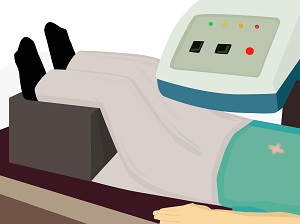
DXA Scanning of Lumbar Spine – L-Spine DXA Anatomy, Regions of Interest, Radiographic Appearance, patient instructions, Patient Positioning, ROI Placement, L-Spine DXA Common Problems, poor bone edge detection, nonremovable artifacts, L-Spine DXA variant anatomy, Fractures or Pathology
DXA Scanning of Proximal Femur – Femur / Hip DXA Anatomy, Regions of Interest, Radiographic Appearance, patient instructions, Patient Positioning, ROI Placement, Common Problems, poor bone edge detection, nonremovable artifacts, variant anatomy, Fractures or Pathology
DXA Scanning of Forearm – Forearm DXA Anatomy, Regions of Interest, Radiographic Appearance, patient instructions, Patient Positioning, ROI Placement, Common Problems, poor bone edge detection, nonremovable artifacts, variant anatomy, Fractures or Pathology
ARRT®* STRUCTURED EDUCATION CREDIT DISTRIBUTION FOR THIS COURSE.
| DISCIPLINE | CATEGORY & SUBCATEGORIES | CE CREDITS PROVIDED |
|---|---|---|
| BD | Patient Care – Patient Bone Health, Care, and Radiation Principles | 10.00 |
| BD | Procedures – DXA Scanning | 6.00 |
| BD | Image Production – Equipment Operation and Quality Control | 6.00 |
| CT | Safety – Radiation Safety and Dose | 2.00 |
| CT | Procedures – Head, Spine, and Musculoskeletal | 2.00 |
| RAD | Procedures – Extremity Procedures | 2.00 |
| RAD | Safety – Radiation Physics and Radiobiology | 1.50 |
| RAD | Safety – Radiation Safety | 1.50 |
| THR | Safety – Radiation Physics and Radiobiology | 1.50 |
| THR | Safety – Radiation Protection, Equipment Operation, and Quality Assurance | 1.50 |
| RA | Safety – Patient Safety, Radiation Protection, and Equipment Operation | 5.00 |
| RA | Procedures – Musculoskeletal and Endocrine Sections | 3.00 |
| NMT | Safety – Radiation Physics, Radiobiology, and Regulations | 3.00 |
| MRI | Procedures – Musculoskeletal | 2.00 |
