Radiographic Positioning of the Lumbar Spine
This article discusses radiographic positioning of the lumbar spine for the Radiologic Technologist (X-Ray Tech).
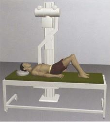
Radiographic positioning guide.


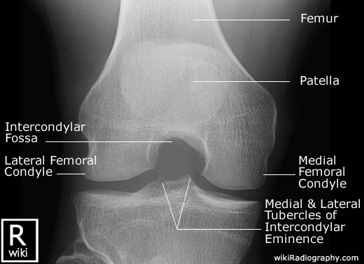
This is an article for Radiologic Technologists (X-Ray Techs) about radiographic positioning of the knee in AP projections.
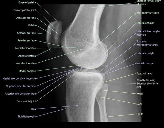
This article is intended for the Radiologic Technologist (X-Ray Tech). It discusses radiographic positioning of the knee in PA projections.
This article for Radiologic Technologists (X-Ray Techs) discusses radiographic positioning of the knee in lateral views.